
Chest digital image cardiothoracic ratio measuring method

A technology of digital imaging and measurement methods, applied in image enhancement, image analysis, image data processing and other directions, can solve the problems of large ratio error, slow speed, prolonged waiting time of patients in queue, etc., to achieve improved stability and adaptability, The effect of saving measurement time

Active Publication Date: 2017-07-28
心医国际数字医疗系统(大连)有限公司
View PDF3 Cites 9 Cited by
  • Summary
  • Abstract
  • Description
  • Claims
  • Application Information

AI Technical Summary

Problems solved by technology

[0003] Among them, the cardiothoracic ratio (refers to the ratio of the transverse diameter of the heart (the sum of the maximum distance from the left and right heart margins to the midline of the body) on the X-ray film to the transverse diameter of the thorax (the inner diameter of the chest through the level of the right diaphragm)) is used to judge whether a person's heart is hypertrophy One of the indicators; the current status is that doctors either use measuring tools to measure on-site on X-ray films or use the auxiliary diagnostic tools of imaging software to measure and finally calculate the ratio of the two; these two measurement methods are doctors positioning through the eyes, and then Manually measuring the diameter, for pixel-level images, the ratio error obtained by visual inspection is obviously a bit large, which is not accurate enough for the diagnosis of the patient's condition, and this measurement method is relatively slow, which prolongs the time for doctors to see a doctor, and also prolongs the time for later patients. waiting time in line
[0004] For machine measurement, image processing is particularly important. The existing technology relies heavily on image input quality, and the algorithm stability and adaptability are not high.

Method used


Image

  • Chest digital image cardiothoracic ratio measuring method
  • Chest digital image cardiothoracic ratio measuring method
  • Chest digital image cardiothoracic ratio measuring method

Examples


Embodiment

[0035] Existing cardiothoracic ratio calculations are mainly performed manually by doctors. In this process, doctors are relatively subjective and rely on their eyes for positioning. For pixel-level images, the error of doctors' visual inspection is obviously somewhat large. For modern hospitals, each doctor needs to diagnose many patients every day. The current manual calculation method is obviously slow, which affects the progress and the waiting time is too long. The four-corner information of the image does not save the result of the cardiothoracic ratio. For the situation where it is necessary to refer to the previous image data and read the film again, every time the image is taken out, it must be recalculated. This repetitive work increases the workload of the doctor. However, in the existing image processing process, conventional gray-scale threshold segmentation is difficult to completely segment the lung lobes, and the multi-mode matching method cannot meet the requir...


PUM


Abstract

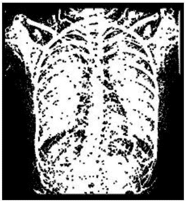
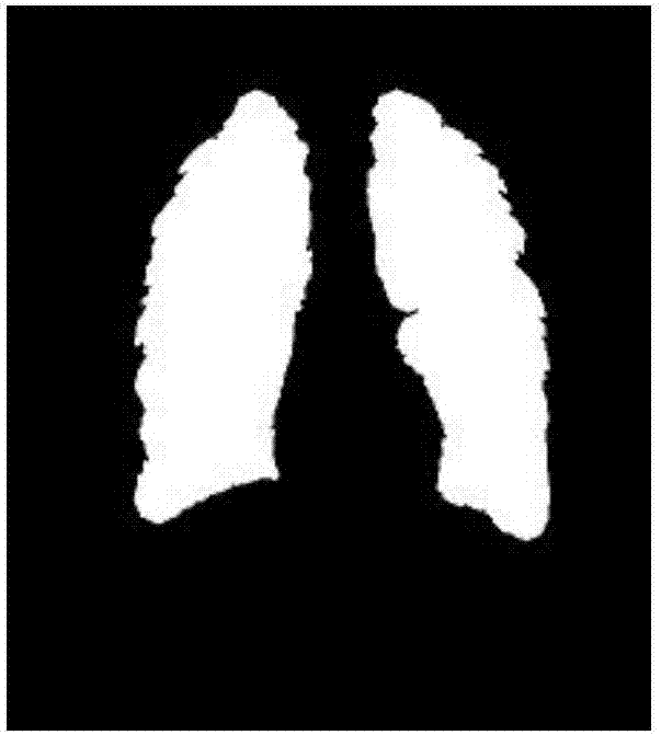
The invention provides a chest digital image cardiothoracic ratio measuring method and belongs to the field of measurement and image processing. The method is mainly characterized by carrying out image preprocessing before segmentation, and based on the preprocessing technology, obtaining different skeleton images through two successive selections of different Gaussian error parameters; on the basis of constructing a reasonable filtering function, constructing a structure operator suitable for rib width for top-hat operation; finally, carrying out primary threshold processing on the images obtained after top-hat operation to obtain a primary lung lobe binary image; adjusting error parameters of the filtering function to obtain a thorax outer edge skeleton image; filling gaps between outer edge skeletons and lung lobes to obtain a lung lobe binary image; and with a rectangular region wherein the binary image locates serving as a region of interest, re-calculating the gray threshold of the region of interest, and obtaining a final lung lobe binary image. The method allows a doctor to automatically calculate the cardiothoracic ratio while reading a film, and give an accurate positioning point.

Description

technical field [0001] The invention belongs to the field of measurement and image processing, and relates to a method for measuring cardiothoracic ratio of chest digital images. Background technique [0002] Compared with hospitals ten years ago, there are many more information-based devices in hospitals today. The emergence of these devices aims to help doctors make auxiliary diagnoses, which greatly facilitates doctors' work. Reading CR, DR and other X-ray films to get the patient's condition is what cardiologists must do every day. [0003] Among them, the cardiothoracic ratio (referring to the ratio of the transverse diameter of the heart (the sum of the maximum distance from the left and right heart borders to the midline of the body) on the X-ray film to the transverse diameter of the thorax (the inner diameter of the chest through the level of the right diaphragm)) is used to judge whether a person's heart is hypertrophy One of the indicators; the current status is ...

Claims


Application Information

Patent Timeline
IPC IPC(8): G06T7/62G06T7/11G06T7/136G06T7/155G06T7/13G06T7/00
CPCG06T7/0012G06T2207/10116G06T2207/20036G06T2207/30048G06T2207/30061
Inventor 王兴维邰从越郭晓红刘龙李小英
Owner 心医国际数字医疗系统(大连)有限公司
Learn More