
Method for constructing engineering arterial blood vessel in vivo by taking melt-spinning fiber as skeleton

A melt spinning, arterial blood vessel technology, applied in the direction of prosthesis, single-component polyester rayon, single-component copolyester rayon, etc. The effect of good mechanical strength, good biocompatibility

Inactive Publication Date: 2017-05-31
NANKAI UNIV
View PDF0 Cites 2 Cited by
  • Summary
  • Abstract
  • Description
  • Claims
  • Application Information

AI Technical Summary

Problems solved by technology

However, traditional biological tubes (made by implanting medical silicone tubes in the abdominal cavity or subcutaneously of animals) have thin walls, weak mechanical properties, and cannot support the tubular shape, which limits the application of biological tubes. Therefore, it is imminent to improve the mechanical properties of traditional biological tubes.

Method used

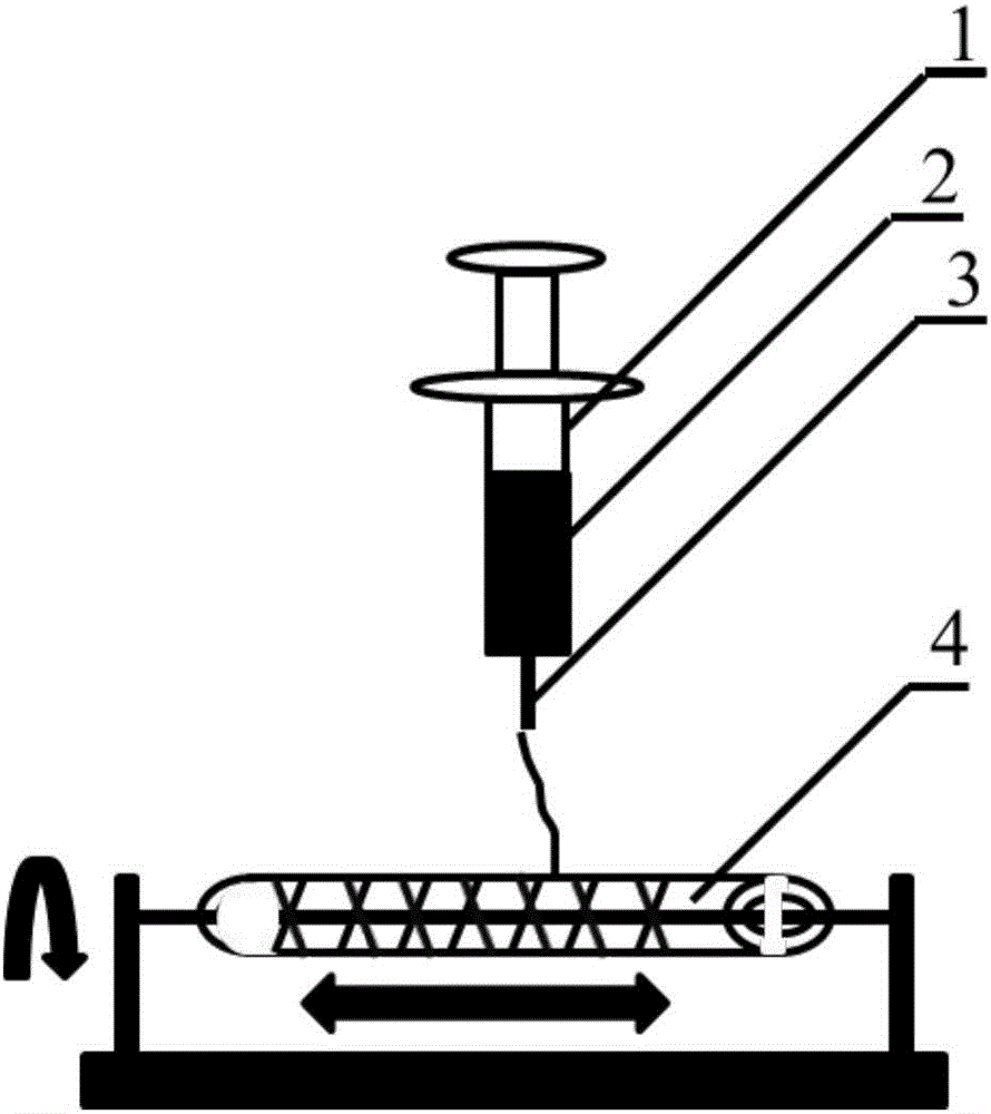
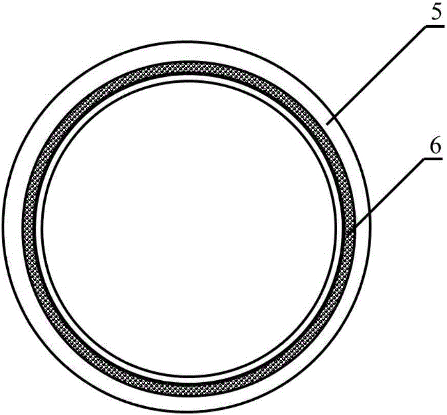
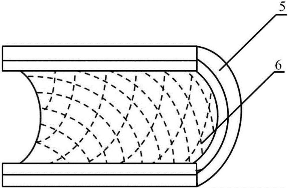

Image

  • Method for constructing engineering arterial blood vessel in vivo by taking melt-spinning fiber as skeleton
  • Method for constructing engineering arterial blood vessel in vivo by taking melt-spinning fiber as skeleton
  • Method for constructing engineering arterial blood vessel in vivo by taking melt-spinning fiber as skeleton

Examples


Embodiment 1

[0023] A method for constructing engineered arteries in vivo using melt-spun fibers as a skeleton, using polycaprolactone (PCL) spun fibers as a skeleton, and subcutaneous rats as a bioreactor for the preparation of engineered arteries in vivo, specific steps as follows:

[0024] 1. Preparation of PCL melt-spun fiber and medical silicone tube composite mandrel

[0025]Weigh 5.0g, with a number average molecular weight of 80000PCL, place it in a closed stainless steel syringe wrapped in a fuser, and heat it at 220°C for 1h. Put a medical silicone tube with an outer diameter of 2mm and an inner diameter of 1mm on a stainless steel rod with a diameter of 1mm and connect it to the rotating motor. The speed is set to 300r / min, and the translation speed is set to 10mm / s. The spinning time was set to 10 min, and the composite mandrel of PCL melt-spun fiber and medical silicone tube with a fiber angle of 45° was prepared. After the spinning is completed, the silicone tube is withdr...

Embodiment 2

[0031] A method for constructing engineered arteries in vivo with melt-spun fibers as a skeleton, using polycaprolactone (PCL) spun fibers as a skeleton, and preparing an in vivo engineered arteries in a rabbit subcutaneous bioreactor, the specific steps are as follows :

[0032] 1. Preparation of PCL melt-spun fiber and medical silicone tube composite mandrel

[0033] Weigh 5.0g, number average molecular weight of 80000PCL, put it in a closed stainless steel syringe wrapped in a fuser, and heat at 220°C for 1h. Put a medical silicone tube with an outer diameter of 2mm and an inner diameter of 1mm on a stainless steel rod with a diameter of 1mm and connect it to the rotating motor. The speed is set to 300r / min, and the translation speed is set to 10mm / s. The spinning time was set to 15 min, and the composite mandrel of PCL melt-spun fiber and medical silicone tube with a fiber angle of 45° was prepared. After the spinning is completed, the silicone tube is withdrawn from th...

Embodiment 3

[0039] A method for constructing engineered arteries in vivo with melt-spun fibers as a skeleton, using polyglycolic acid (PGA) spun fibers as a skeleton, and subcutaneous rats as a bioreactor for the preparation of engineered arteries in vivo, the specific steps are as follows :

[0040] 1. Preparation of PGA melt-spun fiber and medical silicone tube composite mandrel

[0041] Weigh 5.0 g of PGA with a number average molecular weight of 100,000, place it in a closed stainless steel syringe wrapped in a fuser, and heat at 300°C for 1 hour. Put a medical silicone tube with an outer diameter of 2mm and an inner diameter of 1mm on a stainless steel rod with a diameter of 1mm and connect it to the rotating motor. The speed is set to 300r / min, and the translation speed is set to 10mm / s. The spinning time was set to 10 min, and the composite mandrel of PGA melt-spun fiber and medical silicone tube with a fiber angle of 45° was prepared. After the spinning is completed, the silico...


PUM


Abstract

The invention provides a method for constructing engineering arterial blood vessel in vivo by taking melt-spinning fiber as a skeleton. The method comprises the following steps: firstly preparing a melt spinning and medical silica gel tube composite mandrel, and then preparing the in-vivo engineering arterial blood vessel taking melt-spinning fiber as a skeleton by taking subcutaneous part of an animal as a bioreactor; the specific steps are as follows: 1) preparing the melt spinning and medical silica gel tube composite mandrel; 2) preparing the in-vivo engineering arterial blood vessel taking melt-spinning fiber as a skeleton by taking subcutaneous part of an animal as a bioreactor; and 3) performing in-situ transplanting of the in-vivo engineering arterial blood vessel so as to replace lesion blood vessels. The method has the advantages that the in-vivo engineering arterial blood vessel has good biocompatibility, all the components are originated from autologous tissues, and therefore, the artificial blood vessel is non-toxic, free from immunogenicity, and incapable of causing inflammation; due to the existence of melt spinning fiber, the prepared in-vivo engineering blood vessel has good mechanical strength, tenacity and compliance, and is suitable for surgical sewing operation.

Description

technical field [0001] The invention relates to a preparation method of an artificial blood vessel, in particular to a method for constructing an engineered arterial blood vessel in vivo with melt-spun fibers as a skeleton. Background technique [0002] Cardiovascular disease and peripheral vascular disease are among the diseases with the highest morbidity and mortality worldwide. It is estimated that by 2030, there will be as many as 23.3 million deaths related to cardiovascular disease. When the blood vessels are less narrow, treatments include angioplasty and stenting. But for highly narrowed or even blocked blood vessels, vessel bypass surgery is necessary, which has reached about 400,000 cases per year in the United States alone. Autologous blood vessels, the gold standard for vascular grafts, such as the saphenous vein and internal mammary artery, are commonly used in arterial bypass surgery. However, the use of autologous blood vessels requires a second operation, ...

Claims


Application Information

Patent Timeline
Patent Type & Authority Applications(China)
IPC IPC(8): A61L27/50A61L27/38A61L27/18A61L27/02A61L27/24D01F6/62D01F6/84
CPCA61L27/18A61L27/025A61L27/24A61L27/3804A61L27/507D01F6/62D01F6/84
Inventor 王恺张秋影孔德领朱美峰韩晶黄瑞
Owner NANKAI UNIV
Learn More