Preparation of electrochemical biosensor device based on hemoglobin-nano-palladium-graphene composite materials and applied research of electrochemical biosensor device
A biosensor and hemoglobin technology, applied in the field of electrochemical biosensors and chemically modified electrodes, can solve problems affecting direct electrochemical behavior
- Summary
- Abstract
- Description
- Claims
- Application Information
AI Technical Summary
Problems solved by technology
Method used
Image
Examples
Embodiment 1
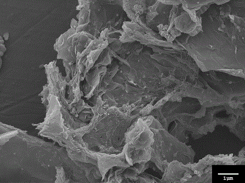
[0028] Scanning Electron Microscopy Characterization of Pd-GR Nanocomposites
[0029] figure 1 Shown is a scanning electron microscope characterization of the Pd-GR nanocomposite at a magnification of 10000. The layered structure is the characteristic morphology of graphene, and palladium nanoparticles are deposited, intercalated, and embedded on the surface and inside of the layered graphene.
Embodiment 2
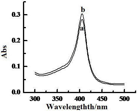
[0031] UV-Vis Absorption Spectroscopy Analysis
[0032] Ultraviolet-visible absorption spectroscopy is a common method for detecting whether the secondary structure of a protein changes. Whether the position of the Soret absorption band of the protein shifts can provide information on the physical structure of the protein. If the protein denatures or its physical structure changes, it will It will cause its absorption band to migrate or disappear. figure 2 The UV-vis absorption spectra of Hb aqueous solution and Pd-GR-Hb mixed solution are presented, the Soret absorption band of Hb in aqueous solution is at 405 nm (curve a) and the Soret absorption band of Hb-Pd-GR mixed solution is 405 nm ( Curve b) is exactly the same, indicating that Hb still maintains the original conformation after being mixed with Pd-GR composite material, and there is no structural change, which further shows that Hb still maintains the original shape in the composite film mixed with Pd-GR composite ma...
Embodiment 3
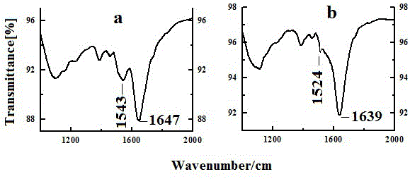
[0034] Infrared Spectroscopy Analysis
[0035] Fourier transform infrared spectroscopy (FT-IR) detects whether the secondary structure of the protein has changed by detecting the changes in the two absorption characteristic bands of amide I and amide II of the protein. The stretching vibration of the C=O bond contained in the peptide structure of the protein causes the amide I (1700-1600 cm -1 ) changes, N-H bond bending vibration and C-N bond stretching vibration caused by amide II (1620-1500 cm -1 ) changes. If the protein is changed or denatured, the two absorption bands of amide I and amide II may be shifted or even disappeared. Such as image 3 The infrared absorption bands of Hb amide I and amide II mixed with Pd-GR composites are shown at 1639 cm -1 and 1524 cm -1 (b), while the absorption band of native Hb is 1647 cm, respectively -1 and 1543 cm -1(a). From the similarity of the infrared spectrum shape, it can be considered that Hb basically maintains its origi...
PUM
 Abstract
Description
Claims
Application Information
 - R&D
- Intellectual Property
- Life Sciences
- Materials
- Tech Scout
- Unparalleled Data Quality
- Higher Quality Content
- 60% Fewer Hallucinations
Browse by: Latest US Patents, China's latest patents, Technical Efficacy Thesaurus, Application Domain, Technology Topic, Popular Technical Reports.
© 2025 PatSnap. All rights reserved.Legal|Privacy policy|Modern Slavery Act Transparency Statement|Sitemap|About US| Contact US: help@patsnap.com


