Addressable sample plate and application thereof in single cell microscopic imaging
A sample plate, single-cell technology, applied in measurement devices, material analysis by optical means, instruments, etc., can solve problems such as difficulty in positioning single cells, and achieve the effect of reducing difficulty
- Summary
- Abstract
- Description
- Claims
- Application Information
AI Technical Summary
Problems solved by technology
Method used
Image

Examples
Embodiment 1
[0043] This example is used to illustrate the preparation and characterization of the addressable sample plate of the present invention.
[0044] (1) Preparation of chromium template
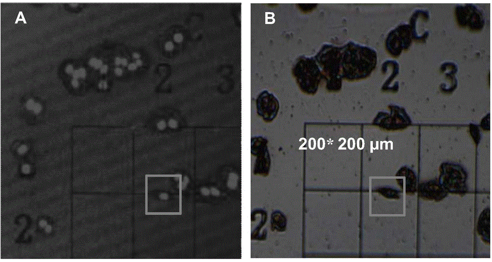
[0045] Using EBL electron beam exposure technology to write as Figure 7 On-board graphics shown (A, B, C, D, 1, 2, 3, 4, 4 × 4 arrays, each cell size is 50 × 50 μm 2 ), and corrode the chromium film to make a chromium template.
[0046] (2) Preparation of sample plate
[0047] The template obtained in the above step (1) is used for ultraviolet exposure to make a patterned photoresist mask. 1×1cm under the protection of the mask 2 ICP silicon etching is performed on the double-sided polished silicon wafer, and the width and depth required for etching are both 3 μm. Wash three times ultrasonically with 5 mL of acetone, and blow dry with nitrogen to obtain a sample plate.
[0048] (3) Characterization of the sample plate
[0049] Observation of the obtained sample plate under a microscope (...
Embodiment 2
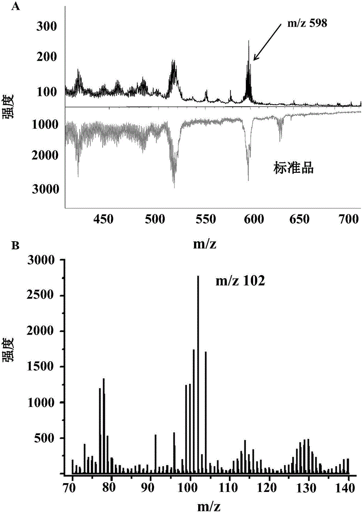
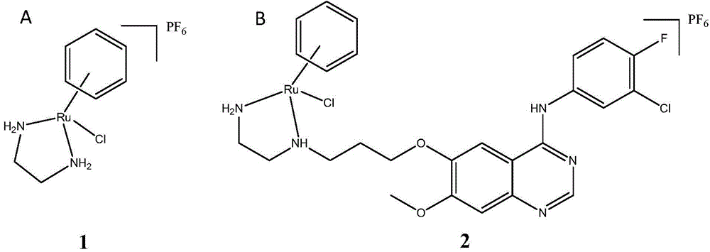
[0051] This embodiment is used to illustrate that the sample plate prepared in Example 1 is used for SIMS-Confocal combined imaging research on ruthenium compound 1 (structural formula such as figure 2 A) Method of spatial distribution in MCF-7 cells.
[0052] (1) Culture and lyophilization of MCF-7 cells
[0053] Place the sample plate in the center of the bottom of the cell culture dish for laser confocal, digested MCF-7 cells in 10 4 cells / cm 2 The concentration was cultured in a petri dish for 24 hours, and the medium containing 100 μM ruthenium compound 1 was added to incubate for 24 hours, the medium was removed, and the culture medium containing 1 μg / mL Hoechst 33342 was incubated in a 37°C incubator for 10 minutes. Remove the medium, and then incubate for 20 min in a 37°C incubator with a culture medium containing 5 μM cell membrane red fluorescent dye. The medium was removed, and the cells were treated with NH at a concentration of 150 mM 4 COOCH 3 (pH 7.4) buff...
Embodiment 3
[0064] This embodiment is used to illustrate that the sample plate prepared in Example 1 is used for SIMS-Confocal combined imaging research on ruthenium compound 2 (structural formula such as figure 2 B) Means of spatial distribution in MCF-7 cells.
[0065] (1) Culture and lyophilization of MCF-7 cells
[0066]Place the sample plate in the center of the bottom of the cell culture dish for laser confocal, digested MCF-7 cells in 10 4 cells / cm 2 The concentration was cultured in a petri dish for 24 hours, and the medium containing 100 μM ruthenium compound 2 was added to incubate for 24 hours, the medium was removed, and the culture medium containing 1 μg / mL Hoechst 33342 was incubated in a 37°C incubator for 10 minutes. Remove the medium, and then incubate for 20 min in a 37°C incubator with a culture medium containing 5 μM / L cell membrane red fluorescent dye. The medium was removed, and the cells were treated with NH at a concentration of 150 mM 4 COOCH 3 (pH 7.4) buff...
PUM
| Property | Measurement | Unit |
|---|---|---|
| diameter | aaaaa | aaaaa |
| width | aaaaa | aaaaa |
| depth | aaaaa | aaaaa |
Abstract
Description
Claims
Application Information
 - R&D
- Intellectual Property
- Life Sciences
- Materials
- Tech Scout
- Unparalleled Data Quality
- Higher Quality Content
- 60% Fewer Hallucinations
Browse by: Latest US Patents, China's latest patents, Technical Efficacy Thesaurus, Application Domain, Technology Topic, Popular Technical Reports.
© 2025 PatSnap. All rights reserved.Legal|Privacy policy|Modern Slavery Act Transparency Statement|Sitemap|About US| Contact US: help@patsnap.com