
OCT-based in-situ three-dimensional printing skin repairing equipment and implementation method thereof

A skin repair and three-dimensional printing technology, applied in the field of biomedical engineering, can solve problems such as limited imaging resolution, long technical measurement time, and large equipment, and meet the requirements of reducing the speed of cell enrichment, instant skin repair, and real-time The effect of monitoring

Active Publication Date: 2016-11-09
REGENOVO BIOTECH
View PDF1 Cites 29 Cited by
  • Summary
  • Abstract
  • Description
  • Claims
  • Application Information

AI Technical Summary

Problems solved by technology

There are two printing methods of 3D bioprinting: in vitro printing and in situ printing in vivo. Currently, most of the 3D bioprinting researches that have been reported use in vitro printing. This method has many limitations in clinical application. First, because the printing materials involve For living cells, it is necessary to ensure a sufficiently high cell survival rate and strictly control the microenvironment of cell growth during the printing process. At the same time, it is also necessary to consider how to maintain tissue growth and metabolism through vascularization and how to regulate subsequent differentiation processes. Technology The difficulty is very high; secondly, the tissues or organs printed out in vitro are rich in fluid biomaterials and have poor mechanical properties, so it is very difficult to transplant and fix them into in vivo tissues under strict aseptic conditions. In addition, printing cultured tissues in vitro The organ takes a long time, and the shape of the defect site may have changed during implantation, which will cause size mismatch problems during transplantation
However, the common imaging technology has the following problems when used for imaging defect skin: the X-ray of micro-computed tomography (micro-CT) has a large ion radiation to the skin tissue, and the imaging contrast is limited ;Magmetic resonance imaging (MRI) technology takes too long to measure, the equipment is huge, and the imaging resolution is limited (mm level), it is difficult to scan and measure skin defects next to the operating table; ultrasound imaging can realize real-time skin scanning, However, its imaging resolution is limited (about 0.5mm), the image speckle noise is large, and the sector scanning method is used, which brings great challenges to the analysis and modeling of skin defects after imaging; confocal microscopy, multiphoton microscopy ( The imaging depth of optical imaging methods such as Multiphoton microscopy (MPM) is limited. For example, the imaging depth of confocal microscopy for highly scattering samples is about 100 μm, and the penetration depth of MPM is only 400-500 μm.
Public reports show that the current digital models based on these imaging technologies can print only 20 layers of cells and only a few hundred microns thick, which is far from meeting actual needs; laser 3D scanning technology can quickly obtain skin defect parts without contact The external contour of the skin, but the microscopic information of the internal tissue structure of the skin cannot be obtained

Method used


Image

  • OCT-based in-situ three-dimensional printing skin repairing equipment and implementation method thereof
  • OCT-based in-situ three-dimensional printing skin repairing equipment and implementation method thereof
  • OCT-based in-situ three-dimensional printing skin repairing equipment and implementation method thereof

Examples


Embodiment Construction

[0041] Below in conjunction with accompanying drawing, the present invention will be further described

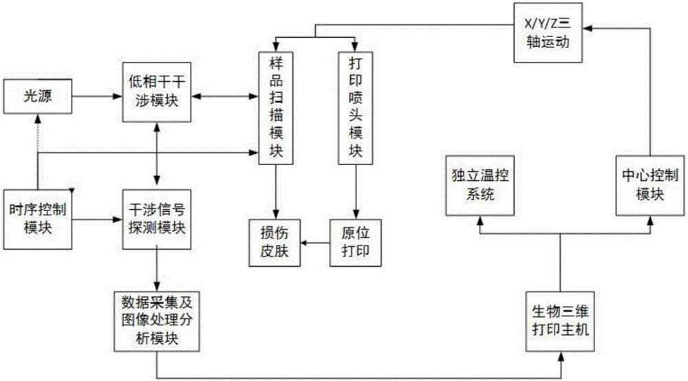
[0042] The in situ three-microbial printing system in vivo includes a 3D bioprinting device based on optical coherence tomography and a 3D bioprinting device with controllable printing parameters. The schematic block diagram is as follows: figure 1 shown.

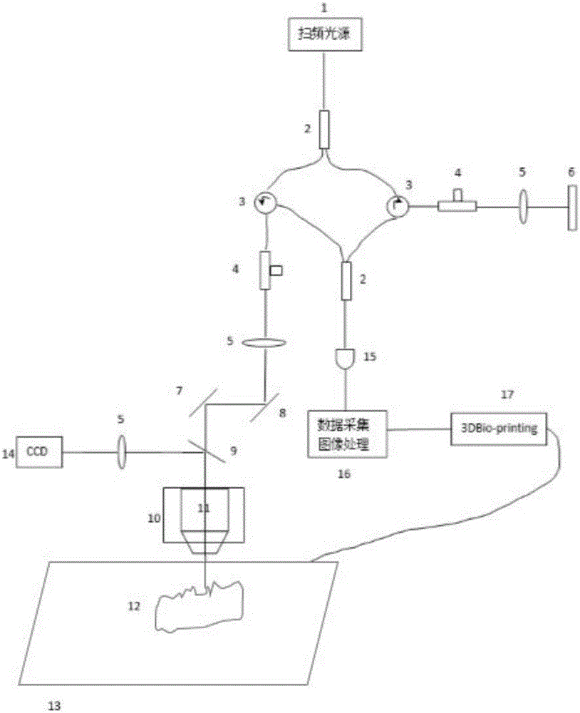
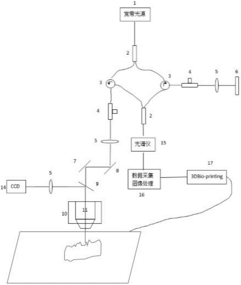
[0043] Figure 2(a) is a detailed view of the in situ 3D bioprinting system based on swept source coherence tomography (SS-OCT), and Figure 2(b) is based on spectral domain coherence tomography (SD-OCT) A detailed view of the in situ 3D bioprinted skin repair system in vivo. The working principle is: the light emitted by light source 1 (Fig. 2(a) is a frequency-sweeping light source, and Fig. 2(b) is a broadband light source) is divided into two paths by a 1×2 fiber coupler 2, one of which is a reference arm, and the light passes through The fiber optic circulator 3, the fiber polarization controller 4 and the collimato...


PUM


Abstract

The invention discloses OCT-based in-situ three-dimensional printing skin repairing equipment and an implementation method thereof. The implementation method comprises the following steps: scanning a skin injury region by adopting an OCT technology to acquire a high-resolution three-dimensional skin injury OCT image; carrying out three-dimensional bionic structural design and modeling on the skin injury part based on the OCT image so as to ensure the reconstruction requirements of skin repair to layered interfaces and vascular networks in tissues; and then, transmitting the modeled skin injury repair model data to a 3D biological printer to carry out model layering and printing, thereby implementing quick, accurate and in-situ repair on the injured part. The OCT-based in-situ three-dimensional printing skin repairing equipment has the advantages of no contact, no injury and real-time imaging, meets the requirement of skin in-situ printing on high-resolution imaging of internal micro-structures, and can acquire distribution and density information of blood vessels in the skin corium layer so as to be convenient for constructing a three-dimensional model closer to real skin structures and functions.

Description

technical field [0001] The invention belongs to the technical field of biomedical engineering, and relates to a three-dimensional printing device based on optical coherence tomography (OCT) for skin repair and its realization method, in particular to an OCT-based principle for skin repair A three-dimensional printing device and its implementation method. Background technique [0002] Three-dimensional (three-dimensional, 3D) printing is a technology based on digital imaging technology to manufacture three-dimensional entities through layered processing and superposition molding, while three-dimensional bio-printing (three-dimensional Bio-printing, 3DBio-printing) is the process of 3D printing On the basis of bio-ink (biocompatible materials, cells, growth factors, etc.) It has great application prospects in solving tissue repair and shortage of donor organs. At present, 3D bioprinting technology is still in its infancy and is developing from printing simple life forms to c...

Claims


Application Information

Patent Timeline
IPC IPC(8): A61B5/107B33Y30/00B33Y50/00A61F2/10
CPCA61B5/1079A61F2/105B33Y30/00B33Y50/00A61B5/00Y02P10/25
Inventor 王玲徐铭恩宋江新杜显斌
Owner REGENOVO BIOTECH
Learn More