Eureka AIR delivers breakthrough ideas for toughest innovation challenges, trusted by R&D personnel around the world.

Biological 3D printing apparatus

A 3D printing and bioprinting technology, applied in metal processing equipment, 3D object support structures, processing and manufacturing, etc., can solve the problems of minimally invasive surgery for tissues and organs that are not applicable, and achieve a wide range of applications, fast curing speed, and simple operation Effect

Active Publication Date: 2016-07-13
南通皋特医疗科技有限公司
View PDF3 Cites 22 Cited by
  • Summary
  • Abstract
  • Description
  • Claims
  • Application Information

AI Technical Summary

Problems solved by technology

[0005] However, the existing bio-3D printing equipment uses a fixed molding platform, and the nozzle can only work in a limited molding space, which has the following disadvantages:
[0006] ①The printing of biological three-dimensional structures such as implants, tissue engineering scaffolds, tissues and organs can only be completed in vitro, and the printing and repair of required tissues or organs cannot be completed in real time in animals or humans
[0007] ②The nozzle can only be moved by three-dimensional motion device, which is not suitable for minimally invasive surgery of various tissues and organs

Method used


Image

  • Biological 3D printing apparatus
  • Biological 3D printing apparatus
  • Biological 3D printing apparatus

Examples


Embodiment

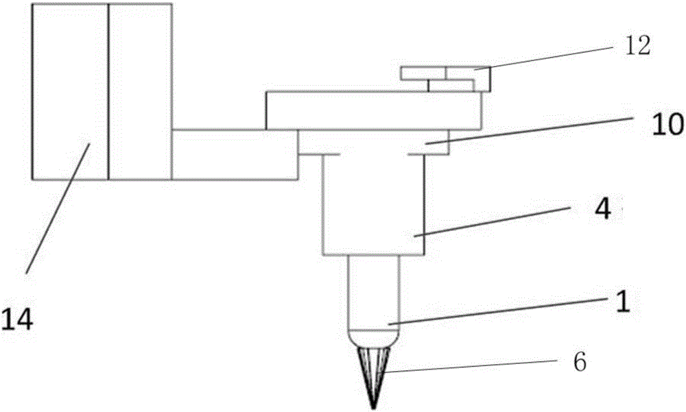
[0036] Such as Figure 1-Figure 3 , This equipment includes printing pen 1, air pump 2, air pressure controller 3, 3D bioprinting pen device 4, printing pen head 6, pen body casing 7, nozzle sleeve 9, connecting piece 10, L-shaped fixing bolt 12, screw hole 13 And printer stand 14;

[0037] The air pump 2, the air pressure controller 3, and the printer frame 14 are connected in sequence. The printer frame is provided with a detachable 3D bioprinting pen device 4, which includes a pen body shell 7 and a pen body shell 7 The printing pen 1 is disassembled. The top of the printing pen 1 is connected to the air pump 2 through a pipe to receive the solution for 3D printing. The bottom end of the printing pen 1 includes a printing pen head 6, and a connecting piece 10 is sleeved on the pen body shell 7. The connecting piece 10 is connected to the printer frame 14 to fix the printing pen 1 on the printer frame 14, wherein the bottom of the pen body shell 7 is provided with a through ho...


PUM


Abstract

The invention provides a biological 3D printing apparatus, comprising a printing frame.A 3D biological printing pen device is disposed on the printing frame and comprises a pen barrel shell and a removable printing pen inside the pen barrel shell, the bottom end of the printing pen includes a printing nib, and a connecting sheet sleeves the pen barrel shell and is connected with the printing frame.An upper end of the printing pen is connected with an air pump hose, a nozzle casing sleeves the pen barrel shell, the top of the nozzle sleeve is provided with a rotary L-shaped fixing bolt, the L-shaped fixing bolt can be rotated to the top end of the printing pen to limit the printing pen, and after the L-shaped fixing bolt is rotated outside the printing pen, the printing pen is available for removal.The biological 3D printing apparatus forms an open space, the printing pen can be removed for handheld operation, and real-time minimally invasive in-situ printing for tissues or organs is facilitated.

Description

Technical field [0001] The invention belongs to the technical field of biological printing in tissue engineering, and particularly relates to a biological 3D printing device. Background technique [0002] Tissue engineering is the application of the principles and methods of engineering and life sciences to prepare biologically active artificial substitutes to maintain, restore or improve part or all of the functions of human tissues and organs. Tissue engineering can be applied to many aspects including bone and cartilage tissue engineering, skin tissue engineering, heart, liver, kidney and other internal organs. [0003] Biological 3D printing is the use of the basic principles and methods of 3D printing discrete / stacked forming, controlled printing of biological materials (including natural biological materials and synthetic biological materials) or cell suspensions to form the required biologically active implants The three-dimensional structure of objects, cells, or artificia...

Claims


Application Information

Patent Timeline
Patent Type & Authority Applications(China)
IPC IPC(8): B29C67/00B33Y30/00A61F2/02
CPCB33Y30/00A61F2/02B33Y70/00B29L2031/753B29K2105/0035A61L27/16B29C64/209B29C64/106A61L27/14A61L27/38B33Y10/00B29C64/264B33Y80/00B33Y50/02B29C64/393
Inventor 蒋青朱莉娅李澜李宗安李客楼杨建飞
Owner 南通皋特医疗科技有限公司
Why Eureka
Eureka Blog
Learn More