
3D kidney model printing method for kidney stone surgical simulation teaching

A technology for surgery simulation and kidney stones, which is applied in the field of 3D kidney model printing in the teaching of kidney stone surgery simulation, which can solve the problems of easily missing anatomical structures, lack of three-dimensional understanding of the patient's kidney and stones, and prone to danger

Inactive Publication Date: 2015-12-02
汤润
View PDF0 Cites 25 Cited by
  • Summary
  • Abstract
  • Description
  • Claims
  • Application Information

AI Technical Summary

Problems solved by technology

[0003] The technical problem to be solved by the present invention is to provide a 3D kidney model printing method for the simulation teaching of kidney stone surgery. Choosing the best puncture point is the difficulty and key point in the operation. At present, doctors can only find the puncture point through preoperative two-dimensional CT images and intraoperative B-ultrasound positioning, lacking a three-dimensional understanding of the patient's kidneys and stones. Wanting to form a three-dimensional figure in the mind through two-dimensional images not only requires long-term training, but also easily misses some small anatomical structures, and dangerous problems are more likely to occur in some difficult and anatomical variations

Method used


Image

  • 3D kidney model printing method for kidney stone surgical simulation teaching
  • 3D kidney model printing method for kidney stone surgical simulation teaching
  • 3D kidney model printing method for kidney stone surgical simulation teaching

Examples


Embodiment Construction

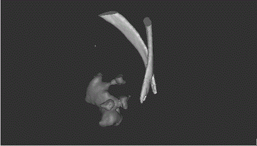
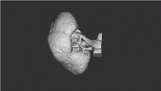
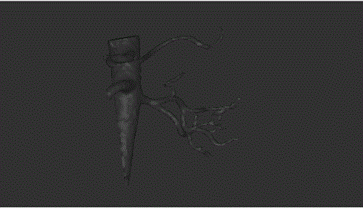
[0021] like figure 1 , 2 , 3, 4, 5, and 6, the present invention uses MIMICS software to obtain a high-density calculus three-dimensional reconstruction model in the plain scan phase of the CTU, obtains a three-dimensional reconstruction model of the renal cortex and arteriovenous vessels through the arteriovenous enhancement phase, Obtain a 3D reconstruction model of the renal pelvis and ureter during the period of enhanced secretion, so as to obtain a 3D reconstruction model of the kidney including the renal cortex, renal medulla, renal artery and vein, renal pelvis and calices, and ureter, and finally combine the 3D reconstruction models of each part Obtain a complete three-dimensional reconstruction model of the kidney; the above method comprises the following steps:

[0022] 1) The patient routinely underwent CT urography. CT urography included plain scan, arteriovenous phase, and excretion phase; In the venous phase, the gray values ​​of the aorta, renal arteriovenous,...


PUM


Abstract

The invention discloses a 3D kidney model printing method for kidney stone surgical simulation teaching. The printing method comprises the following steps: acquiring a high-density stone three-dimensional rebuilt model by utilizing MIMICS software by virtue of a scanning period of CTU, acquiring a three-dimensional rebuilt model of kidney skin and arteriovenous blood vessels by virtue of an arteriovenous enhanced period, acquiring a three-dimensional rebuilt model of pelvis and ureter by virtue of secretion enhanced period of pelvis, acquiring a kidney three-dimensional rebuilt model comprising kidney skin, kidney medulla, kidney artery and vein, pelvis and calices and ureter, and finally combining the three-dimensional rebuilt model of each part to obtain a complete kidney three-dimensional rebuilt model. By adopting the method, the problems that percutaneous nephrolithotomy is used as an effective and safe minimally-invasive surgery, to select an optimum puncture point is difficulty and a key point in the surgical operation, the formation of a three-dimensional image in the brain by virtue of a two-dimensional image needs long-term training and is prone to missing small dissection structures can be effectively solved.

Description

technical field [0001] The invention belongs to the technical field of medical auxiliary equipment, and in particular relates to a 3D kidney model printing method used for the simulation teaching of kidney stone operation. Background technique [0002] Urinary calculus is one of the common diseases in urology, accounting for the first place in the number of urology hospitalizations. Since 1976, Fernstrom et al. first applied nephrolithotomy through the percutaneous puncture and expanded nephrostomy channel to remove stones from the renal pelvis successfully. The gold standard for multiple calculus or calculi. PCNL is an effective and safe minimally invasive surgery, but it has its own special complications. The incidence of intraoperative and postoperative massive bleeding was about 13.7%, and the incidence of colon, spleen, and liver injury was about 0.4%. To maximize the lithotripsy rate and reduce complications has become a hot spot in the development and research of P...

Claims


Application Information

Patent Timeline
IPC IPC(8): A61B19/00G09B23/30B33Y10/00B33Y30/00G06T17/00G06F17/18
Inventor 汤润朱海涛汪洋孙晓青
Owner 汤润
Learn More