Preparation method of rapidly decellularized unifoliate liver biological scaffold

A bioscaffold and decellularization technology, applied in medical science, prostheses, etc., can solve the impact on the quality and stability of decellularized liver bioscaffold preparation, the lack of uniform standards for perfusion pathways and protocols, and the low replantation rate of decellularized tissues and other problems, to achieve the effect of shortening the required time, good effect, and short solution time

Active Publication Date: 2015-11-11
WENZHOU MEDICAL UNIV
View PDF4 Cites 3 Cited by
  • Summary
  • Abstract
  • Description
  • Claims
  • Application Information

AI Technical Summary

Problems solved by technology

[0003] Since 2010, foreign scholars have successfully prepared whole liver decellularized bioscaffolds by perfusion with different detergents[4,7-11], but there is no uniform standard for the perfusion pathway and protocol, which will affect the preparation quality and quality of decellularized liver bioscaffolds. serious impact on stability
However, as a commonly used detergent, the residue of SDS will lead to easy formation of thrombus after tissue transplantation, and the low replantation rate of decellularized tissue during recellularization[13]

Method used


Image

  • Preparation method of rapidly decellularized unifoliate liver biological scaffold
  • Preparation method of rapidly decellularized unifoliate liver biological scaffold
  • Preparation method of rapidly decellularized unifoliate liver biological scaffold

Examples


Embodiment Construction

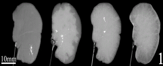
[0019] Obtaining whole liver of SD rats

[0020] Healthy adult SD rats were weighed, anesthetized by intraperitoneal injection of 0.3-0.4ml / 100g10% chloral hydrate, fixed after the effect, opened the abdominal cavity, isolated the inferior vena cava, and injected heparin sodium solution for systemic heparinization. Find the hepatic hilum located below the liver, separate the hepatic artery, bile duct and portal vein, cut off the bile duct, ligate and cut off the beginning of the hepatic artery and portal vein, cut off the hepatic vein at the same time, separate the part of the liver connected to the diaphragm, and remove the liver. One group of livers was treated with perfusion decellularization, and the other group of livers was stored at -80°C as a control group.

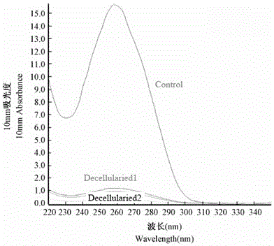
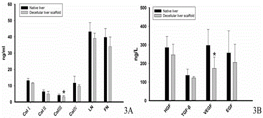
[0021] Preparation of Decellularized Single Lobe Liver Bioscaffolds

[0022] (1) A group of livers were stored at -80°C without further processing as a control group.

[0023] (2) Preparation of decellularized s...


PUM


Abstract

The invention discloses a preparation method of a rapidly decellularized unifoliate liver biological scaffold. An ionic detergent sodium dodecyl sulfonate (SDS) with the problem that thrombus is easy to form after tissue transplantation and the problem of low replantation rate of decellularized tissues in a composite cellularization process is not used; a decellularized left outer-lobe liver biological scaffold is prepared by a weakly alkaline Triton solution perfusion method; due to the addition of NaOH in the Triton solution, the neutral Triton solution is in alkalescence; the cell lysis removal effect is enhanced; the time required for removing cells is shortened; and the optimal concentration and dosage of the NaOH and the Triton solution are adopted, so that the decellularization effect is the best.

Description

technical field [0001] The invention relates to the field of organ tissue decellularization, in particular to a method for preparing a rapid single-lobe liver decellularized biological scaffold. Background technique [0002] Extracellular matrix (extracellular matrix, ECM) is a substance packed between cells, and its components include water, electrolytes, and a small amount of liquid phase components, mainly collagen, mucin, and proteoglycans, which together constitute the microenvironment for cell growth . The local microenvironment of hepatocytes is a variety of cytokines secreted by the extracellular matrix and stromal cells, including hepatocyte growth factor (HGF), vascular endothelial growth factor (VEGF), basic fibroblast growth factor (bFGF), epidermal growth factor (EGF), transforming growth factor (TGF-β), and platelet-derived growth factor (PDGF). Among them, ECM not only provides a three-dimensional spatial structure for the growth of intrahepatic cells, but a...

Claims


Application Information

Patent Timeline
Patent Type & Authority Applications(China)
IPC IPC(8): A61L27/36
Inventor 王志斌梅劲陈纳孟卓张建色余雅玲王志翊黎婷
Owner WENZHOU MEDICAL UNIV
Why Eureka
Try Eureka