
Cell-biological scaffold complex and 3D printing forming method thereof

A bio-scaffold and 3D printing technology, applied in prosthetics, medical science, additive processing, etc., can solve the problems of difficult adhesion of cells, large pore size, and inability to form pores, so as to improve the loss of cell suspension, mechanical The effect of good performance and controllable shape profile

Active Publication Date: 2015-09-09
深圳尤尼智康医疗科技有限公司
View PDF4 Cites 43 Cited by
  • Summary
  • Abstract
  • Description
  • Claims
  • Application Information

AI Technical Summary

Problems solved by technology

[0003] The existing 3D printing technology has realized the controllable manufacture of the shape of the scaffold, which meets the requirements of the preparation of the gradient scaffold in terms of process principle, but the pore size constructed by this technology is large, and the pores below 100 μm cannot be formed, and it cannot provide a microstructure for cell growth. environment, there is a problem that cells are not easy to adhere to
The forming material of 3D printing technology is generally a single material or a composite material artificially mixed in advance, but there are still great limitations in forming scaffolds with material gradient characteristics
However, the scaffold made of fibers ejected by electrospinning technology is similar to the disordered non-woven fabric, so it is difficult to precisely control the shape of the scaffold.
It is not difficult to see that the current single process method cannot achieve multi-scale forming, so it cannot meet the macroscopic contour forming requirements and microscopic pore forming requirements of bone scaffolds at the same time.

Method used


Image

  • Cell-biological scaffold complex and 3D printing forming method thereof
  • Cell-biological scaffold complex and 3D printing forming method thereof
  • Cell-biological scaffold complex and 3D printing forming method thereof

Examples


Embodiment 1

[0040] 1, the preparation of PCL-nHA composite material, the mass ratio of described PCL and nHA is 4:1:

[0041] Weigh 4g of polycaprolactone PCL and add it to 36g of dichloromethane solution, stir on a magnetic stirrer until fully dissolved, add 1g of nHA hydroxyapatite powder, stir on a magnetic stirrer until the solution is uniform, vacuumize and degas , placed in a fume hood until the organic solvent is completely volatilized, and set aside.

[0042] 2 Preparation of collagen-chitosan electrospinning solution:

[0043] Chitosan and collagen were dissolved in deionized water successively at a mass ratio of 1:2 to prepare an electrospinning solution with a mass fraction of 8%.

[0044] 3 Preparation of bone cell suspension:

[0045] Transfer the culture flask containing osteoblasts to the ultra-clean workbench, remove the old culture medium, add trypsin to digest for 1 minute, remove the trypsin, add DMEM / F12 cell culture medium containing 20% ​​serum to terminate the dig...


PUM

PropertyMeasurementUnit
Apertureaaaaaaaaaa
Apertureaaaaaaaaaa

Abstract

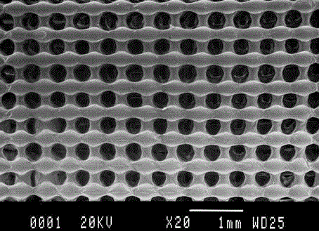
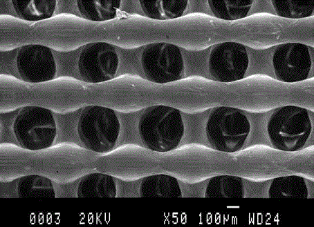
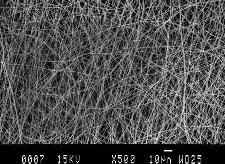
The invention provides a cell-biological scaffold complex and a 3D printing forming method thereof. The cell biological scaffold complex comprises a biological scaffold with an actual deficit condition similar to the tissues and organs of patients, a nanometer fiber layer printed on the biological scaffold, capable of simulating natural extracellular matrixes, and beneficial for the adhesion, reproduction and growth of cells, and cell suspension printed on the nanometer fiber layer. According to the actual deficit condition of the tissues and organs of patients, the cell-biological scaffold complex needed by patients can be precisely printed by adopting the bioprinting forming method.

Description

technical field [0001] The invention belongs to the field of biotechnology, in particular to a cell-biological scaffold complex and a 3D printing forming method thereof. Background technique [0002] Tissue defect repair and organ transplantation are one of the greatest breakthroughs in the history of medicine in the 20th century. At present, there are nearly 1 million patients in the world who were once shrouded in the shadow of death and gained a new life through organ transplantation, but the number of patients who died due to lack of donor organs and organ transplantation is as high as tens of millions, and patients who need tissue defect repair It is as high as hundreds of millions of people, so some people say that the 21st century is the century of tissue repair and organ transplantation. The emergence of 3D printing technology has brought hope to millions of patients who need tissue repair and organ transplantation. [0003] The existing 3D printing technology has ...

Claims


Application Information

Patent Timeline
IPC IPC(8): A61L27/38A61L27/42A61L27/48A61L27/56A61L27/54A61L27/58B29C67/00B33Y10/00
Inventor 王红胡海龙张楠施敏超
Owner 深圳尤尼智康医疗科技有限公司
Learn More