A CT Image Reconstruction Method for Expanding Field of View

A CT image, expanding field of view technology, applied in the field of biomedical imaging, can solve problems such as incompleteness, and achieve the effect of good image fusion

Active Publication Date: 2017-06-06
北京锐视康科技发展有限公司
View PDF6 Cites 0 Cited by
  • Summary
  • Abstract
  • Description
  • Claims
  • Application Information

AI Technical Summary

Problems solved by technology

[0011] In order to overcome the deficiencies of the above-mentioned prior art, the present invention provides a CT image reconstruction method for expanding the field of view, which utilizes PET image information to carry out supplementary reconstruction of CT images outside the CT field of view (ie, the maximum physical imaging diameter), thereby overcoming Image loss and image artifacts caused by incomplete data problems can be used for image fusion and image correction in PET / CT equipment
The method provided by the present invention can not only utilize the PET image contour information, but also fully utilize the edge information of the CT tomographic projection space image (called a sino map), and perform a good supplementary reconstruction of the missing CT image's morphology and material properties, preferably effectively overcome the image loss and image artifacts caused by the incomplete data problem; the present invention utilizes a method of assigning value curve by curve to sino images with missing CT image edge information in CT projection space, and utilizes the edge information of CT projection space to the greatest extent. Numerical information can realize the continuity condition of the data flow at the edge of the CT projection space, avoid the prior assumption of a single medium, and better solve the technical difficulties such as the continuity of the projection space data flow in the existing methods

Method used


Image

  • A CT Image Reconstruction Method for Expanding Field of View

Examples


Embodiment Construction

[0031] Below in conjunction with accompanying drawing, further describe the present invention through embodiment, but do not limit the scope of the present invention in any way.

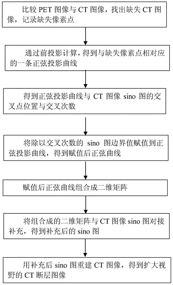
[0032] In PET / CT clinical scanning, when a part of the obese patient's body (such as the shoulder) exceeds the physical field of view of the CT, the CT image obtained by using the PET / CT clinical scanning cannot synthesize the correct PET attenuation correction coefficient, which may Causes artifacts to appear on PET images, interfering with doctors' diagnoses. The present invention supplements and reconstructs the missing CT images by using the obtained PET image information, not only utilizes the PET image contour information, but also makes full use of the edge information of the CT projection space, and performs a very detailed analysis of the shape and material properties of the missing CT images. Good supplementary reconstruction can better overcome the missing images and image artifacts caused...


PUM


Abstract

The invention discloses a CT (computed tomography) image reconstruction method for expanding vision. The CT image reconstruction method for expanding the vision comprises the following steps of performing supplementary reconstruction on a CT image out of a CT vision diameter by using a one-by-one curve assignment method through PET (positron emission tomography) image information to obtain a vision-expanded CT image; finding out and recording pixel points without CT images; obtaining a sine projection curve corresponding to the pixel points; obtaining positions of intersection points of the sine projection curve and a sino graph cut-off boundary of the CT image and the intersection frequency of the sine projection curve and the sino graph cut-off boundary of the CT image; combining the sine projection curve into a two-dimensional matrix after all points on the sine projection curve are assigned; performing butt joint supplementing on the two-dimensional matrix and a sino graph detector array of the CT image to obtain a supplemented sino graph; and reconstructing the CT image to obtain a vision-expanded CT cross-sectional image. By the CT image reconstruction method for expanding the vision, image missing and image artifacts which are caused by incomplete data can be avoided, and the CT image reconstruction method can be used for fusing and correcting images in PET / CT equipment.

Description

technical field [0001] The invention belongs to the technical field of biomedical imaging and relates to PET / CT equipment imaging, in particular to a CT image reconstruction method for expanding the field of view, which can be used for image fusion and image correction in PET / CT equipment. Background technique [0002] CT is the abbreviation of Computed Tomography. CT is a physical examination of the human body using X-rays. PET (Peter) is the abbreviation of Positron Emission Tomography, which is a molecular imaging device for functional metabolic imaging. [0003] The full name of PET-CT is positron emission tomography and X-ray computed tomography imaging system, which can provide anatomical imaging and functional imaging at the same time. It is the most ideal combination in current imaging diagnosis technology, especially in the diagnosis and staging of tumors. , efficacy evaluation and other aspects play an important role. [0004] The so-called PET / CT refers to the ...

Claims


Application Information

Patent Timeline
Patent Type & Authority Patents(China)
IPC IPC(8): G06T11/00A61B6/03
Inventor 刘力井上慎一李真曾海宁
Owner 北京锐视康科技发展有限公司
Why Eureka
Try Eureka