
A method for isolating tumor stem cells

A technology of tumor stem cells and tumor cells, which is applied in the direction of tumor/cancer cells, animal cells, vertebrate cells, etc., can solve the problems of not determining TSC-specific surface markers, difficult to effectively separate TSCs, and no unified screening of surface markers, etc., to achieve The effect of good homogeneity, low cost and simple method

Active Publication Date: 2017-01-18
OSINGLAY BIO PHARM CO LTD +1
View PDF2 Cites 0 Cited by
  • Summary
  • Abstract
  • Description
  • Claims
  • Application Information

AI Technical Summary

Problems solved by technology

[0004] Although there are more and more evidences supporting the TSC theory, there are still many problems in the study of TSC: first, not all tumors have been isolated from TSC; second, the technology of TSC isolation and identification is still not perfect; There is no defined TSC-specific surface marker; fourth, the origin, occurrence, and molecular and cellular mechanisms of TSC have not been thoroughly studied, and there are still many major issues to be resolved
It seems that there is a group of cells in the mesenchymal population that can withstand external physical pressure and have stronger self-renewal and differentiation capabilities, and TSC is a type of tumor cells with stronger self-renewal and differentiation capabilities, but each There is no uniform screening surface marker for different types of TSC, and its source is far more than mesenchymal type cells, including a large number of epithelial, neural, lymphoid, blood and other types of tumors, and it is difficult to effectively separate them by using a single unified molecular marker for sorting Various types of TSC

Method used


Image

  • A method for isolating tumor stem cells
  • A method for isolating tumor stem cells
  • A method for isolating tumor stem cells

Examples


Embodiment 1

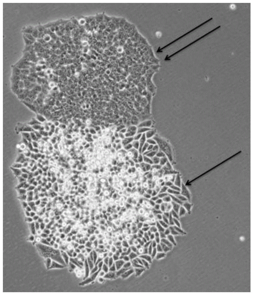
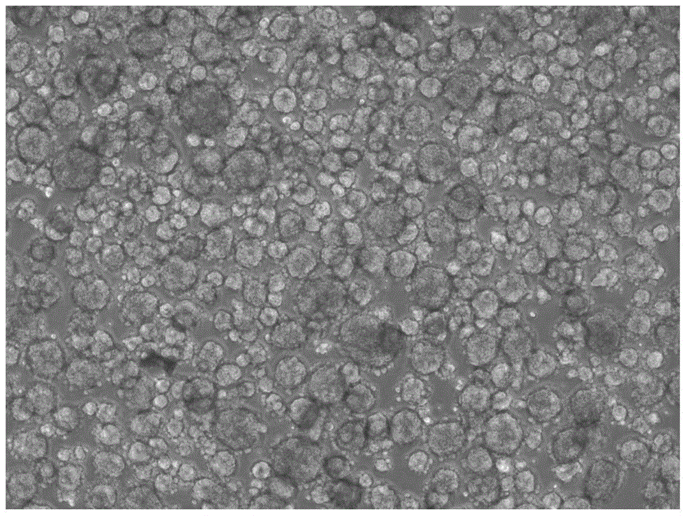
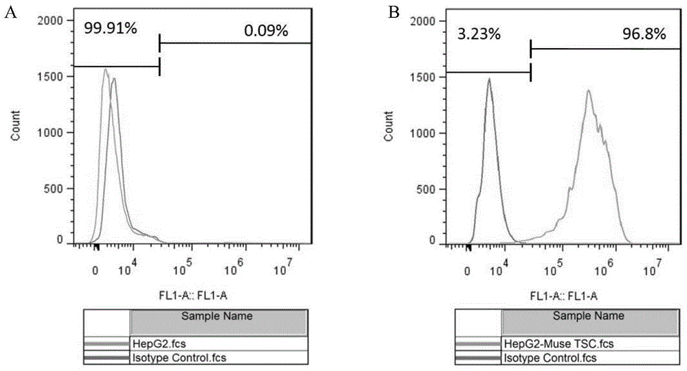
[0082] Example 1 Isolation method of HepG2 liver tumor stem cells

[0083] 1) Digest HepG2 adherent hepatoma cells with 0.25% trypsin solution at 37°C for 2-5 minutes to single cells, terminate with medium containing serum, centrifuge at 200g / min for 5min, and collect the precipitated cells; (here Steps can use other digestive enzymes instead of trypsin solution, such as TrypLE, Accutase, etc., the digestion time, digestion temperature, and centrifugation time are related to different types of tumor cells);

[0084] 2) Add 0.1% type I collagenase solution (diluted with basal medium DMEM / F12) to the collected cells above, every 1×10 6 Add 1mL enzyme solution to each cell and incubate at 37°C for 6h to simulate the digestive enzyme stress state; (in this step, other digestive enzymes can be used instead of 0.1% type I collagenase solution, such as 0.02-0.3% (w / v) type IV Collagenase solution, 0.1~3U / ml Dispase solution, 0.01~0.3% (w / v) trypsin solution, 0.01~1% (w / v) TrypLE enz...

Embodiment 2

[0105] Example 2 Isolation method of Hep3B liver tumor stem cells

[0106] 1) Use 0.25% trypsin solution to digest Hep3B adherent hepatoma cells at 37°C for 2-5 minutes to form a single cell, stop with medium containing serum, centrifuge at 200g / min for 5min, and collect the precipitated cells;

[0107] 2) Add 0.1% type I collagenase solution (diluted with basal medium DMEM / F12) to the collected cells above, every 1×10 6 Add 1mL enzyme solution to each cell and incubate at 37°C for 6h to simulate the stress state of digestive enzymes;

[0108] 3) Move the above-incubated cells to 4°C and incubate for 12-16 hours to simulate low temperature and digestive enzyme stress state;

[0109] 4) Wash the incubated cells with PBS buffer, mix well, centrifuge at 200g / min for 5min, discard the supernatant, wash away the enzyme solution, add DMEM / F12 culture medium to the cells precipitated in the centrifuge tube until the centrifuge tube is full , cover the cap of the centrifuge tube, se...

Embodiment 3

[0117] Example 3 Isolation method of glial tumor stem cells

[0118] 1) Digest adherent glioma cells U251 with 0.25% trypsin solution at 37°C for 4 minutes to single cells, terminate with medium containing serum, centrifuge at 200g / min for 5min, and collect the precipitated cells;

[0119] 2) Add 0.1% type I collagenase solution (prepared from DMEM culture medium) to the collected cells above, every 1×10 6 Add 1mL enzyme solution to each cell and incubate at 37°C for 6h to simulate the stress state of digestive enzymes;

[0120] 3) Move the above-incubated cells to 4°C and incubate for 12-16 hours to simulate low temperature and digestive enzyme stress state;

[0121] 4) Wash the incubated cells with PBS buffer, mix well, centrifuge at 200g / min for 5min, discard the supernatant, wash off the enzyme solution, add DMEM culture medium to the precipitated cells in the centrifuge tube until the centrifuge tube is full, cover Cap the centrifuge tube, seal the bottle with a parafil...


PUM


Abstract

The invention discloses a separating method of tumor stem cells. The separating method comprises the following steps: digesting tumor cells into single cells, then, adding the single cells into a digestive enzyme liquid to hatch for several hours; then, adding the hatched cells into a cell culture fluid containing blood serum to perform adherence growth; after the cells are adhered, transferring the adhered cells into a tumor stem cell TSC culture medium to culture; and after the cells grow all over, repeating the operation for many times, performing microsphere culture on the obtained cells to finally obtain the microspheres which are the tumor stem cells. According to the separating method of the tumor stem cells disclosed by the invention, various digestive enzymes or other physical stress conditions are combined to stimulate various tumor cells, so that TSC in different types can be stably obtained. The MuseTSC separated by the separating method disclosed by the invention has extremely strong stem characteristic and good homogeneity, and is stable, and therefore, the method can be used for enabling the obtaining of the TSC in different types to be standard, relatively simple and economical, and suitable for constructing a TSC base on a large scale to meet the working needs of the conventional tumor biomedicine basic research, the clinical research, the medicine development, and the like.

Description

technical field [0001] The invention belongs to the technical field of tumor stem cells, and relates to a method for separating tumor stem cells. Background technique [0002] In recent years, a large number of reports have found that there are a very small number of cells in the tumor, which are extremely malignant. When the patient undergoes radiotherapy and chemotherapy, the large tumor mass disappears immediately. However, these very small number of cells can still survive, and PET-CT cannot detect them. These cells are cancer stem cells (tumor stem cells, tumor stem cells, TSC; or cancer stem cells, cancer stem cells, CSC; tumor initiating cells , tumor initiating cell, TIC; cancer initiating cell, cancer initiating cell, CIC), many mainstream views today believe that tumor stem cells enriched by radiotherapy and chemotherapy are the main reason for the failure of traditional treatment methods. In 2006, the American Association for Cancer Research defined TSC as: cells...

Claims


Application Information

Patent Timeline
Patent Type & Authority Patents(China)
IPC IPC(8): C12N5/095
Inventor 王淋立陈月花宋立兵
Owner OSINGLAY BIO PHARM CO LTD
Learn More