
In-vivo drug testing system based on nanoparticle modified hollow-core optical fiber

A technology of hollow-core optical fiber and nano-particles, which is applied in the direction of measuring devices, material analysis through optical means, instruments, etc., can solve the problems of affecting signal recording and weak spectral signals, and achieve the effect of no need for marking

Inactive Publication Date: 2015-01-07
SHANGHAI UNIV
View PDF4 Cites 4 Cited by
  • Summary
  • Abstract
  • Description
  • Claims
  • Application Information

AI Technical Summary

Problems solved by technology

At present, measurement and analysis have been carried out directly under the spectrometer, but for in vivo research and analysis, because the Raman spectrum signal is weak, and then collected by optical fiber, the spectral signal is even weaker and affects the signal recording, so signal enhancement is required

Method used


Image

  • In-vivo drug testing system based on nanoparticle modified hollow-core optical fiber
  • In-vivo drug testing system based on nanoparticle modified hollow-core optical fiber
  • In-vivo drug testing system based on nanoparticle modified hollow-core optical fiber

Examples


Embodiment 1

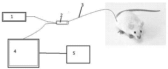
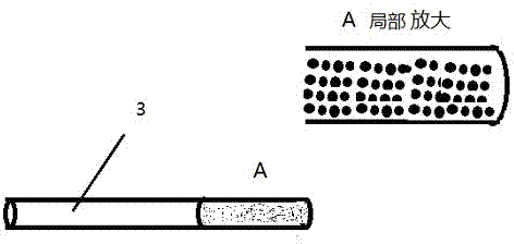
[0019] see figure 1 ~ image 3 , an in vivo drug detection system based on nanoparticle-modified hollow-core optical fiber, including a spectrometer (4), a hollow-core optical fiber (3), a laser light source (1), a lens combination optical path (2) and a computer (5), characterized in that The hollow-core optical fiber (4) is a hollow-core optical fiber modified with nanoparticles; the laser light source (1) is connected to the hollow-core optical fiber (3) through the output end of the lens combination optical path (2), and the hollow-core optical fiber (3) is modified with nanoparticles One end of the lens is inserted into the body; the other output end of the lens combination optical path (2) is connected to the spectrometer (4), and the spectrometer is connected to the computer (5); the laser excites the drug molecules in the body, and the generated optical signal information passes through the hollow-core optical fiber (3) To the lens combination optical path (2), throu...

Embodiment 2

[0022] This embodiment is basically the same as Embodiment 1, and the special feature is: the inner surface of the nanoparticle-modified hollow-core optical fiber is decorated with nanoparticles, and the nanoparticles are gold, silver, copper, or gold-shell silver core or Silver core and gold shell, or nanoparticles or films whose surface is gold, silver or copper. The hollow-core fiber (3) includes a photonic crystal fiber, or a common hollow-core fiber.

Embodiment 3

[0024] This embodiment is basically the same as Embodiment 2, and the special feature is

[0025] The spectrometer (4) uses a Renishaw Invia Confocal Raman Microscope confocal microscope system, the hollow-core fiber (3) uses an ordinary hollow-core fiber, and the nanoparticles modified at one end of the hollow-core fiber (3) are gold nanoparticles of about 50 nm The combined optical path (2) of the particles and the lens adopts an optical path with a filter, a trap and a reflective lens, and the computer (5) is an ordinary computer such as a Lenovo desktop computer. For example, the following test is done: inject ambroxol hydrochloride injection into the intraperitoneal cavity of the mouse, and half an hour later detect the Raman spectrum in the tail vein of the mouse through the hollow-core optical fiber (3). The characteristic peak of the Raman spectrum of ambroxol hydrochloride was detected, and through spectral analysis, it was shown that the mouse blood contained ambroxo...


PUM


Abstract

The invention relates to an in-vivo drug testing system based on a nanoparticle modified hollow-core optical fiber. The system comprises a spectrograph, the hollow-core optical fiber, a laser light source, a lens combination optical path and a computer, wherein the hollow-core optical fiber is the nanoparticle modified hollow-core optical fiber; the laser light source is connected with the hollow-core optical fiber by the output end of the lens combination optical path; one end, modified by nanoparticles, of the hollow-core optical fiber is inserted into a body; the other output end of the lens combination optical path is connected with the spectrograph and the spectrograph is connected with the computer; laser is used for stimulating in-vivo drug molecules to generate optical signal information; then the optical signal information is transmitted to the lens combination optical path through the hollow-core optical fiber and then is transmitted to the Raman spectrograph by the lens combination optical path; and content change information of drugs is judged by analyzing a spectrum through the computer. The system can be used for in-vivo drug testing of animals, human bodies and the like, and provides technical supports for individualized medicines, scientific researches and drug development.

Description

technical field [0001] The invention relates to an in vivo drug analysis system based on nanoparticle-modified hollow-core optical fiber, which uses special optical fiber for biomedical detection and analysis, and provides technical support for current drug development and future personalized medical treatment. Background technique [0002] In vivo drug analysis is an important part of biomedicine and new drug pharmacokinetic research, and is closely related to pharmacokinetic research, clinical pharmacology research and biopharmaceutical research. At present, the analysis methods of drugs in vivo mainly include chromatographic analysis, immunoassay, isotope labeling, and microbial determination. Although these methods are commonly used, in general, these analytical methods are tedious, time-consuming and costly. [0003] With the development of Raman spectroscopy detection methods, Raman spectroscopy detection samples can be directly analyzed and measured without pretrea...

Claims


Application Information

Patent Timeline
IPC IPC(8): G01N21/65G01N21/01
Inventor 陈振宜王廷云商亚娜庞拂飞陈娜刘书朋
Owner SHANGHAI UNIV
Learn More