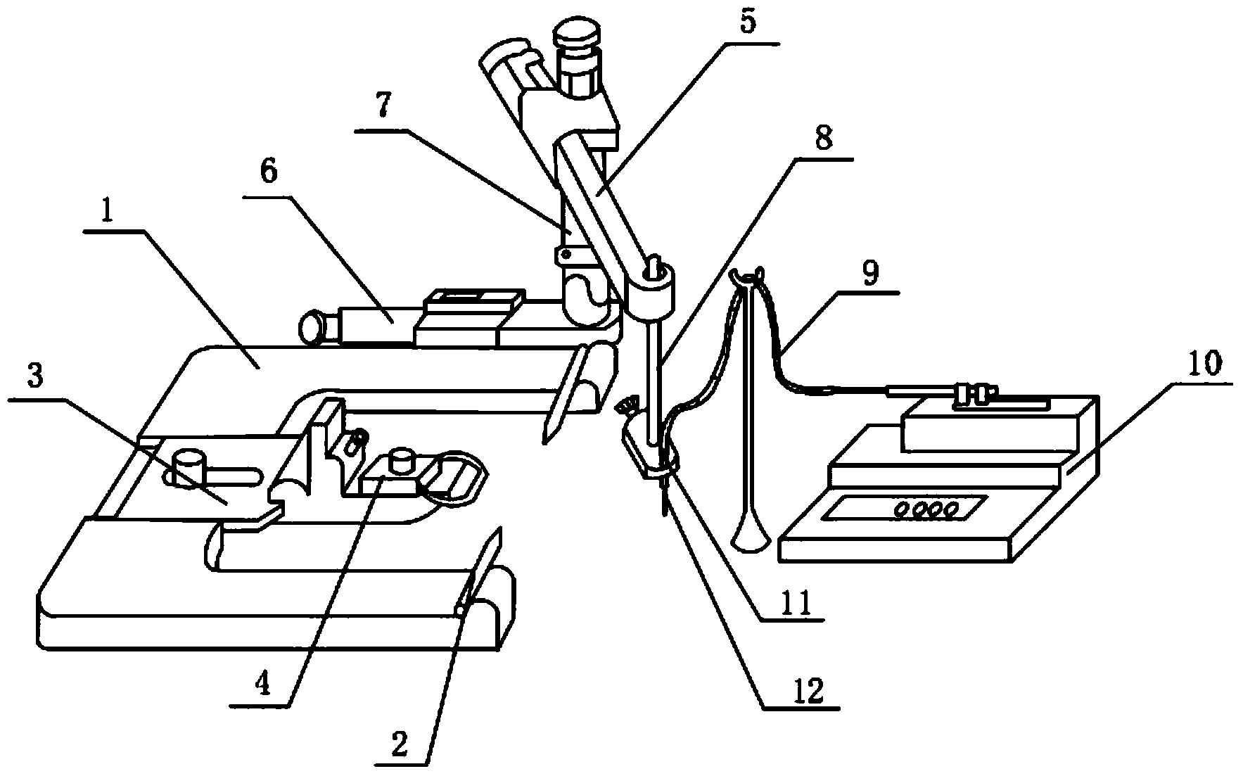
Visual minimally-invasive intracranial dosing device
An internal drug delivery, U-shaped technology, applied in the field of visual minimally invasive intracranial drug delivery device, can solve the problems of inaccuracy, damage to the brain, easy infection, etc., and achieve the effects of accurate positioning, minor damage, and accurate quantitative
- Summary
- Abstract
- Description
- Claims
- Application Information
AI Technical Summary
Problems solved by technology
Method used
Image
Examples
Embodiment 1
[0013] Such as figure 1 , figure 2 As shown, a visible minimally invasive intracranial drug delivery device includes a U-shaped base 1, an ear bar 2 is provided on both sides of the open end of the U-shaped base 1, and an adapter 3 is provided at the closed end of the U-shaped base 1. An incisor clip 4 is provided at the front end of the adapter 3, an X arm 5, a Y arm 6 and a Z arm 7 are provided on the side wall of the U-shaped base 1, and a holder 8 is provided on the X arm 5, wherein: also includes a guide Liquid tube 9, a micro injection pump 10 is installed at one end of the catheter tube 9, a fixed needle 11 is installed at the other end of the catheter tube 9, a capillary quartz tube 12 is installed on the fixed needle 11, and outside the capillary quartz tube 12 The surface is marked with a scale 13, and the outer surface of the catheter 9 is also marked with a scale 13. The catheter 9, the fixed needle 11 and the capillary quartz tube 12 are connected, and the inner...
Embodiment 2
[0015] Medial septum of rat basal ganglia (medial septum, MS):
[0016] To inject 1 μl of drug (such as pontamine blue) into the medial septum of the basal ganglia of an adult SD rat:
[0017] First fill the minimally invasive administration needle with physiological saline, connect the end of the catheter to the micro-injection pump, first suck 0.5 μl of air, and then suck 1 μl of pentamine sky blue. Adult SD rats were anesthetized with chloral hydrate or urethane, fixed on a stereotaxic apparatus 6, and the skull was exposed after hair clipping and disinfection. After drilling the skull 0.7mm behind the anterior bregma and 2mm laterally, slowly insert the fixed minimally invasive drug delivery needle 6mm into the brain. Inclined 18°. The administration was performed by adjusting the administration rate of the microsyringe pump to 0.1 μl / min. see results image 3 :exist image 3 In , the darker area in the middle and lower part of the figure is the medial septum of the r...
Embodiment 3
[0019] Administration of rat hippocampus
[0020] To inject 1 μl of drug (such as pontamine sky blue) into the hippocampal nuclei of an adult SD rat:
[0021] First fill the minimally invasive administration needle with physiological saline, connect the tail end to the micro-injection pump, first suck 0.5 μl of air, and then suck 1 μl of pentamine sky blue. Adult SD rats were anesthetized with chloral hydrate or urethane, fixed on a stereotaxic apparatus, and the skull was exposed after hair clipping and disinfection. After the skull was drilled 3.0mm behind the anterior bregma and 2.2mm laterally, the fixed minimally invasive drug delivery needle was slowly inserted 3.0mm into the cranium. The administration was performed by adjusting the administration rate of the microsyringe pump to 0.1 μl / min. see results Figure 4 :exist Figure 4 In , the brighter area in the lower part of the figure is the hippocampus of the rat. The capillary quartz tube 12 is a dark thin line, a...
PUM
| Property | Measurement | Unit |
|---|---|---|
| The inside diameter of | aaaaa | aaaaa |
| Outer diameter | aaaaa | aaaaa |
Abstract
Description
Claims
Application Information
 - R&D
- Intellectual Property
- Life Sciences
- Materials
- Tech Scout
- Unparalleled Data Quality
- Higher Quality Content
- 60% Fewer Hallucinations
Browse by: Latest US Patents, China's latest patents, Technical Efficacy Thesaurus, Application Domain, Technology Topic, Popular Technical Reports.
© 2025 PatSnap. All rights reserved.Legal|Privacy policy|Modern Slavery Act Transparency Statement|Sitemap|About US| Contact US: help@patsnap.com


