Method and device for establishment of Mycobacterium tuberculosis naturally infected non-human primate model
A primate and non-human technology, applied in animal houses, medical raw materials derived from bacteria, animal husbandry, etc., can solve the problems that cannot be simulated as latent infection
- Summary
- Abstract
- Description
- Claims
- Application Information
AI Technical Summary
Problems solved by technology
Method used
Image
Examples
preparation example Construction
[0055] 1. Preparation of Mycobacterium tuberculosis
[0056] The strain used to infect animals is Mycobacterium tuberculosis H37Rv strain, take 1×10 7 CFU can cause half of the infected mice to die in about 2 weeks (determined by pre-testing) Mycobacterium tuberculosis H37Rv strain preserved on a slant, use a sterile inoculation loop to scrape a little bacteria lawn on the slant, inoculate it on a freshly improved On the slant of Roche's medium, culture at 37°C for 3 weeks. Using a sterile culture ring, scrape the bacterial lawn on the inclined surface, weigh it accurately and grind it in a sterile mortar, add sterile saline at 10-20 mg / mL, and grind it while adding until it is completely dispersed and uniform Cloudy bacteria. Use a quantitative sterile pipette to distribute the bacterial suspension into 1.5mL cryopreservation tubes, 1mL per tube, put them into a marked freezer box, and store them in a -80°C refrigerator as a strain preservation tube. .
[0057] The bacter...
Embodiment 1
[0092] [Example 1] Fiberoptic bronchoscopic infection of Chinese macaques
[0093] 1.1 Anesthesia
[0094] 2 experimental monkeys WNP1 and WNP2 were fasted for 8-12 hours before anesthesia. 15 minutes before anesthesia, intramuscular injection of atropine 0.04 mg / kg body weight and intramuscular injection of ketamine hydrochloride 10 mg / kg body weight gave anesthesia. Afterwards, the animals were placed on the operating table.
[0095] 1.2 Animal infection
[0096] After the animal is anesthetized, lie supine on the operating table, open the mouth with a gag, expose the epiglottis with a laryngoscope, and anesthetize the epiglottis with 1% tetracaine. Operate according to the instructions of the fiber bronchoscope (OLYMPUS BF TYPE XP60, outer diameter 2.8mm), spray 1% lidocaine into the nostrils on both sides of the animal with a sprayer, slowly insert the fiber bronchoscope through the nasal passage of the animal, and when it reaches the epiglottis , slowly inject 1mL of 1...
Embodiment 2
[0099] [Example 2] Mycobacterium tuberculosis infects Chinese rhesus monkeys by natural route—aerosol
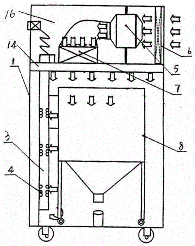
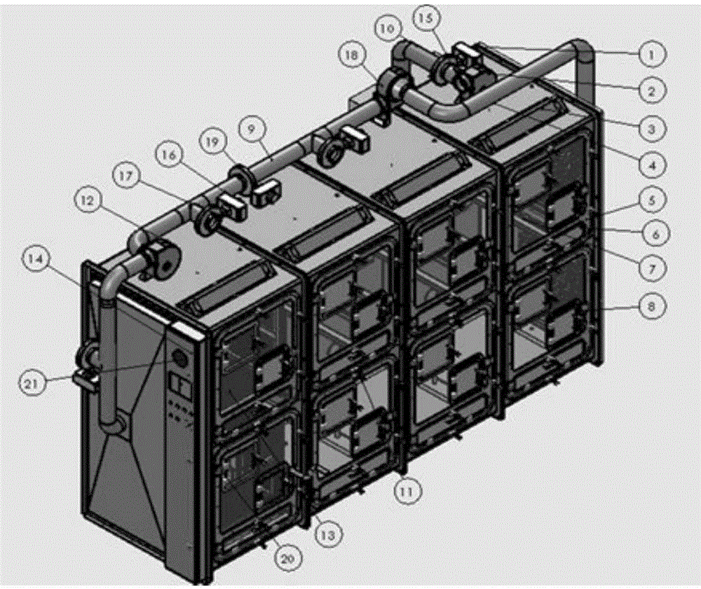
[0100] WPN1 and WPN2 rhesus monkeys that were artificially infected (direct inoculation of Mtb strain into the lungs by fiberoptic bronchoscopy) were placed in an airflow self-circulation combined negative pressure isolator on the day after the artificial infection (see Figure 7 ), and then put 4 healthy monkeys WPN3, WPN4, WPN5, WPN6 into the combined negative pressure isolator, start the airflow self-circulation system, so that the healthy monkeys can repeatedly contact the air from the infected monkey cage, and fresh air can be supplied as required , Control the air supply and exhaust through the PLC control unit, and adjust the air volume. After exposure, clinical symptoms such as appetite and cough were observed every day; various tests were arranged according to Table 3.
PUM
 Abstract
Description
Claims
Application Information
 - Generate Ideas
- Intellectual Property
- Life Sciences
- Materials
- Tech Scout
- Unparalleled Data Quality
- Higher Quality Content
- 60% Fewer Hallucinations
Browse by: Latest US Patents, China's latest patents, Technical Efficacy Thesaurus, Application Domain, Technology Topic, Popular Technical Reports.
© 2025 PatSnap. All rights reserved.Legal|Privacy policy|Modern Slavery Act Transparency Statement|Sitemap|About US| Contact US: help@patsnap.com


