Fluorescent in-situ hybridization method for detecting plant rhizosphere microbe

A fluorescent in situ hybridization and microbial technology, which is applied in biochemical equipment and methods, microbial measurement/testing, fluorescence/phosphorescence, etc., can solve the problems of difficult to find target microorganisms, interfere with microbial observation, and difficult observation, so as to avoid spontaneous fluorescent effect

Active Publication Date: 2013-03-27
RES CENT FOR ECO ENVIRONMENTAL SCI THE CHINESE ACAD OF SCI
View PDF2 Cites 0 Cited by
  • Summary
  • Abstract
  • Description
  • Claims
  • Application Information

AI Technical Summary

Problems solved by technology

The first aspect and the biggest difficulty is that the plant root itself usually has strong autofluorescence
The best time for observation is within the first 24 hours after the collection of plant samples. Afterwards, the gradually enhanced autofluorescence of the plant itself seriously interferes with the observation of microorganisms. However, it is difficult to guarantee timely detection after collection of plant samples in actual operation.
The second aspect is that the sparse distribution of microorganisms in the rhizosphere makes it difficult to find the target microorganisms in the process of microscope observation, and as time goes by, the autofluorescence phenomenon of the root itself gradually strengthens, making observation more difficult

Method used


Image

  • Fluorescent in-situ hybridization method for detecting plant rhizosphere microbe
  • Fluorescent in-situ hybridization method for detecting plant rhizosphere microbe
  • Fluorescent in-situ hybridization method for detecting plant rhizosphere microbe

Examples


Embodiment 1

[0039] Embodiment 1, adopt the method of the present invention to carry out fluorescence in situ hybridization

[0040] 1. Take the root tissue of a Juncus acutiflorus from the wetland. After sampling for 48 hours, wash it with deionized water to remove excess wetland sediment and soil residue attached to the surface; then cut the root tissue into about 2cm in length root fragments.

[0041] 2. Soak the root fragments obtained in step 1 in 2-5ml (whichever is sufficient to completely submerge the root fragments) solution A for 12 hours of fixation.

[0042] The solution A is obtained by mixing 3 parts by volume of fixative solution and 1 part by volume of PBS buffer solution.

[0043] The preparation method of fixative solution is as follows (taking 100ml volume as an example): (1) Take 66ml of ultrapure water and heat it to 60°C in a water bath; (2) Add 4g of paraformaldehyde to the water, the mixture will become turbid; (3) Slowly add 2mol / L NaOH aqueous solution to the mi...

Embodiment 2

[0060] Embodiment 2, adopt the method of the present invention to carry out fluorescence in situ hybridization

[0061] 1. Same as step 1 in Example 1.

[0062] 2. Same as step 2 of embodiment 1.

[0063] 3. Adopt 0.0001mol / L tetrasodium pyrophosphate aqueous solution, incubate in 20 ℃ water bath for 15 minutes, others are the same as step 3 of embodiment 1.

[0064] 4. Same as step 4 in Example 1.

[0065] 5. Same as Step 5 of Example 1.

[0066] 6. Same as step 6 of embodiment 1.

[0067] 7. Same as Step 7 of Example 1.

[0068] 8. Same as step 8 in Example 1.

[0069] 9. Same as step 9 in Example 1.

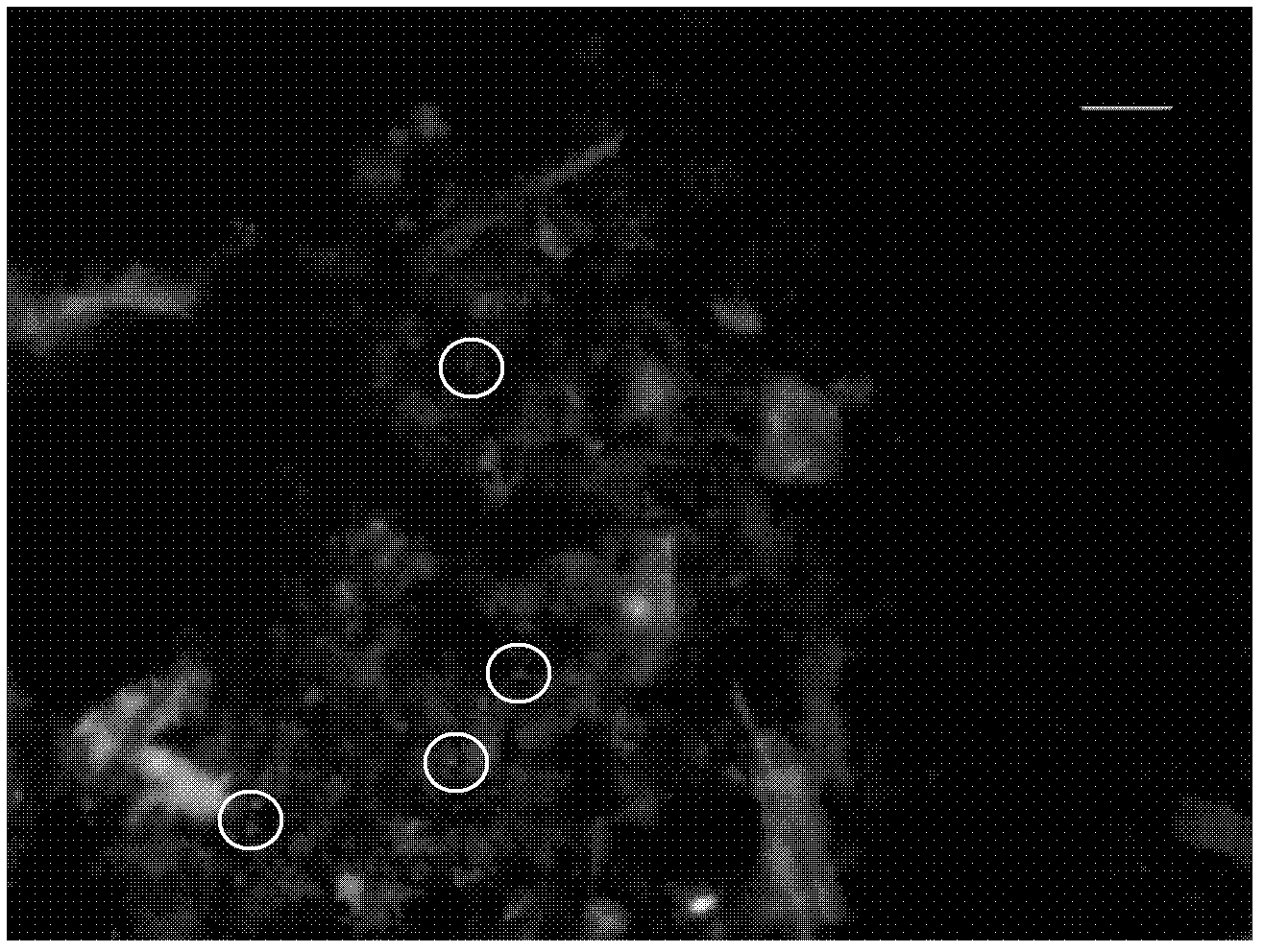
[0070] See figure 2 . The target microorganisms with fluorescence can be clearly observed.

Embodiment 3

[0071] Embodiment 3, adopt the method of the present invention to carry out fluorescence in situ hybridization

[0072] 1. Same as step 1 in Example 1.

[0073] 2. Same as step 2 of embodiment 1.

[0074] 3. Adopt 0.1 mol / L tetrasodium pyrophosphate aqueous solution, incubate in 30° C. water bath for 60 minutes, and other steps are the same as step 3 of embodiment 1.

[0075] 4. Same as step 4 in Example 1.

[0076] 5. Same as Step 5 of Example 1.

[0077] 6. Same as step 6 of embodiment 1.

[0078] 7. Same as Step 7 of Example 1.

[0079] 8. Same as step 8 in Example 1.

[0080] 9. Same as step 9 in Example 1.

[0081] See image 3 . The target microorganisms with fluorescence can be clearly observed.


PUM


Abstract

The invention discloses a fluorescent in-situ hybridization method for detecting plant rhizosphere microbe. The method comprises the following steps of 1) plant rhizosphere fragment fixing; and 2) fluorescent in-situ hybridization; the method is characterized in that a step A is provided between the step 1) and the step 2): the plant rhizosphere fragment completed in the step 1) is processed by 0.0001-0.1mol/L of tetrasodium pyrophosphate aqueous solution. The inventor of the present invention found that the tetrasodium pyrophosphate aqueous solution enables dissociation phenomenon of plant rhizosphere and surface microbe. On this base, a current FISH method is improved, the step of processing by the tetrasodium pyrophosphate aqueous solution is increased between the two steps of fixing by the current FISH mode and the fluorescent in-situ hybridization. The fluorescent in-situ hybridization method for detecting plant rhizosphere microbe solves the problems of the autofluorescence phenomenon of plant, and less content and difficult observation of rhizosphere microbe adhered on the rhizosphere in plant rhizosphere FISH observation.

Description

technical field [0001] The invention relates to a fluorescence in situ hybridization method for detecting plant rhizosphere microorganisms. Background technique [0002] Rhizosphere microbes are microorganisms that grow and reproduce in the soil directly affected by plant roots. The types include bacteria, actinomycetes, fungi, algae, and protozoa. ten times. They are in an interbiotic relationship with plants, interacting with plant roots and promoting each other. According to the coexistence form of plant roots and rhizosphere microorganisms, it can be divided into various forms, the most common ones are leguminous plants and corresponding rhizobia, ectomycorrhizae, endomycorrhizae, and pathogenic bacteria complexes. [0003] In recent years, with the rapid development of metagenomics and molecular ecology technologies, microscopic observations combined with microbial fluorescence in situ hybridization (FISH) have become a powerful tool for understanding rhizosphere micr...

Claims


Application Information

Patent Timeline
Patent Type & Authority Applications(China)
IPC IPC(8): C12Q1/68C12Q1/04G01N21/64
Inventor 祝贵兵王雨尹澄清
Owner RES CENT FOR ECO ENVIRONMENTAL SCI THE CHINESE ACAD OF SCI
Why Eureka
Try Eureka