


Quick Research
Generate reliable direction feasibility study reports for your R&D in just a few steps.

Technical Q&A
Discover and master advanced knowledge NOW. Basics, ideas, possibilities, all at once.

Find Solutions
As an expert in R&D theories, this can generate solutions to your technical problems instantly.

Evaluate Feasibility
Analyze your overall solution with one click, know your potential R&D risks in advance.

Monitor Landscape
Get weekly tech updates, stay abreast of the latest tech innovations and key insights.
Detection method for biological efficacy of umbilical cord mesenchymal stem cell
A stem cell biology, stem cell technology, applied in the field of detection of the biological efficacy of umbilical cord mesenchymal stem cells, can solve problems such as the lack of defined cell quality cell treatment effects
- Summary
- Abstract
- Description
- Claims
- Application Information
AI Technical Summary
Problems solved by technology
Method used
Image


Examples
Embodiment 1
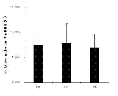
[0022] Example 1 Detection of galectin-3 expression at mRNA level
[0023] Three batches of human umbilical cord mesenchymal stem cells (hUC-MSC) were cultured in a row. After cultured to the P4 generation, they were digested and centrifuged separately. The total RNA of the cells was extracted with an RNA extraction kit (product of Invitrogen), and after reversed to cDNA, galectin-3 The primers were subjected to conventional PCR amplification (forward primer: 5'-CCAAAGAGGGAATGATGTTGCC-3', reverse primer: 5'-TGATTGTACTGCAACAAGTGAGC-3'), and agarose electrophoresis analysis (results) Figure 1A ); Select the same batch of P2~P4 generation cells to extract RNA and reverse it to cDNA, use real-time qPCR (real-time quantitative PCR) analysis, the primer sequence is the same as PCR, and the real-time qPCR result is the target product 2 -ΔCt Analysis (see results Figure 1B ). It can be seen from the results that hUC-MSC expresses galectin-3 at the mRNA level, and there is no significant...
Embodiment 2
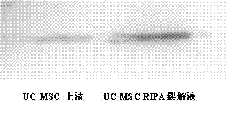
[0024] Example 2 Western blot detection of galectin-3 protein expression
[0025] Cultivate human umbilical cord mesenchymal stem cells with a culture system of 10ml, and collect the supernatant for use after culturing for 48 hours; after cell digestion, centrifuge and count, add 1ml of RIPA reagent (cell lysate, Sigma) for every 1 million cells to lyse the cells. The cell supernatant and RIPA lysate were simultaneously subjected to SDS-PAGE electrophoresis, and the electrophoretic bands were transferred to PVDF membrane by semi-dry method for Western blot analysis. The primary antibody was goat anti-galectin-3 antibody (R&D), and the secondary antibody Rabbit anti-goat IgG (Abcam), see the results figure 2 . It can be seen from the results that hUC-MSC can express galectin-3 protein, and this protein can be secreted into cell culture medium.
Embodiment 3
[0026] Example 3 ELISA to detect galectin-3 protein expression
[0027] Three batches of human umbilical cord mesenchymal stem cells were cultured continuously, the culture system was 10ml, and the supernatant was collected after 48 hours of culture; the cells were digested at the end of 48 hours, and 1ml of RIPA reagent was added for every 1 million cells to lyse the cells. The cell supernatant and RIPA lysate were tested by ELISA (galectin-3 ELISA kit, Bender) at the same time, the results are as follows image 3 . It further proved that galectin-3 protein can be expressed in hUC-MSC cells and can be secreted outside the cell.
PUM
 Abstract
Description
Claims
Application Information
 - R&D Engineer
- R&D Manager
- IP Professional
- Industry Leading Data Capabilities
- Powerful AI technology
- Patent DNA Extraction
Browse by: Latest US Patents, China's latest patents, Technical Efficacy Thesaurus, Application Domain, Technology Topic, Popular Technical Reports.
© 2024 PatSnap. All rights reserved.Legal|Privacy policy|Modern Slavery Act Transparency Statement|Sitemap|About US| Contact US: help@patsnap.com