Device and method for detecting blood vessel deformation area

A vascular area and deformation technology, applied in the field of medical images, can solve problems such as poor accuracy, deviation, and variable physiological characteristics, and achieve the effect of improving sensitivity and accuracy

Active Publication Date: 2011-03-30
SHENYANG NEUSOFT MEDICAL SYST CO LTD
View PDF5 Cites 17 Cited by
  • Summary
  • Abstract
  • Description
  • Claims
  • Application Information

AI Technical Summary

Problems solved by technology

However, the physiological characteristics of the human body are very complex and changeable, and the deformation of a vessel diameter cannot objectively and comprehensively reflect the degree of deformation of the vessel lumen
However, in the above detection process, when only one vessel diameter parameter is used as the only criterion for judging whether the vessel lumen is deformed, the sensitivity of the detection results is reduced
At the same time, in the above-mentioned diameter measurement method, since there is no clear judgment standard for the reference point of the adjacent end and the far end, the detected deformation part has a large deviation from the real situation, and the accuracy is poor.

Method used


Image

  • Device and method for detecting blood vessel deformation area
  • Device and method for detecting blood vessel deformation area
  • Device and method for detecting blood vessel deformation area

Examples


Embodiment 1

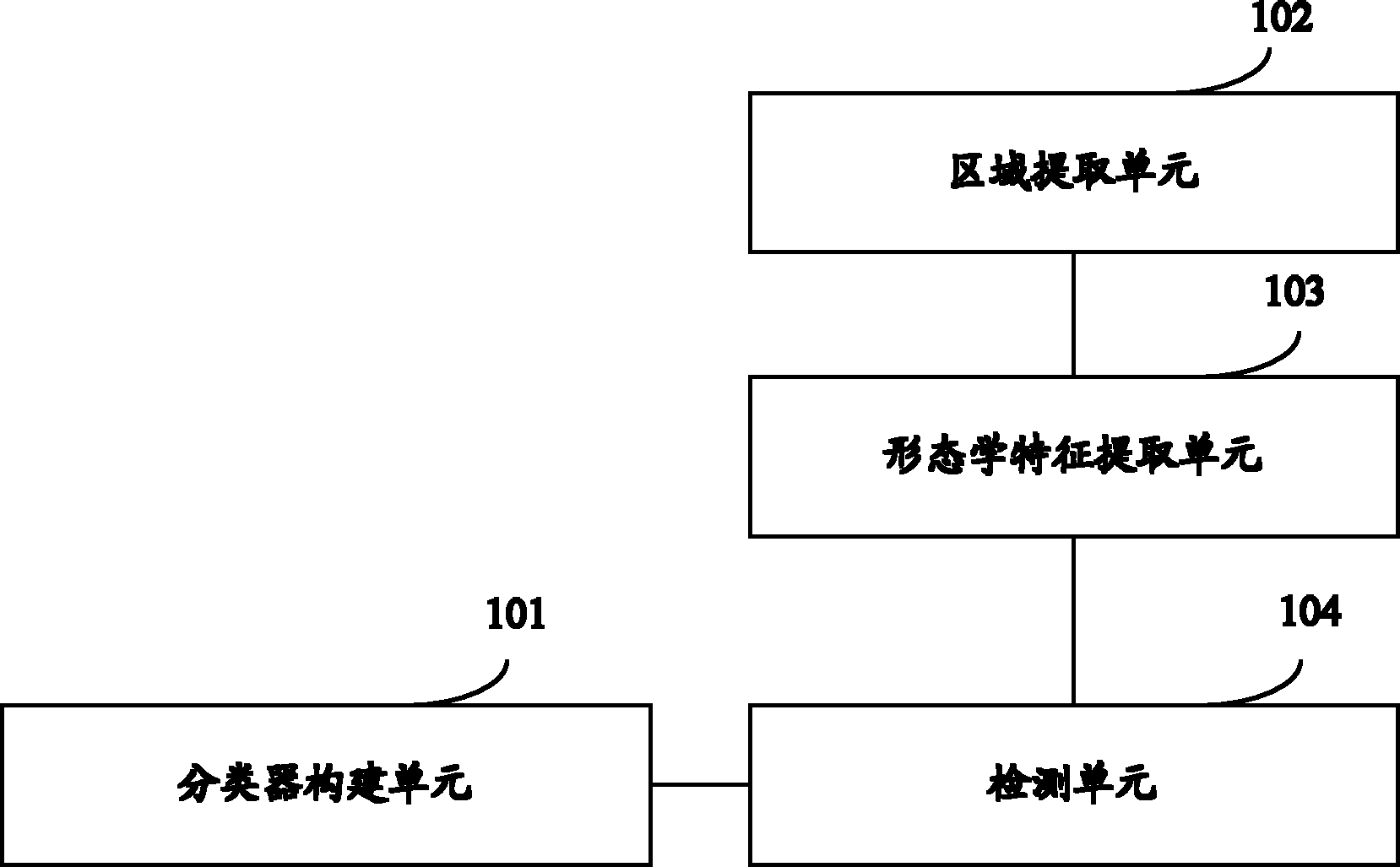
[0044] see figure 1 , which is a schematic structural diagram of an embodiment of a detection device for a vascular deformation region in the present application, the device includes: a classifier construction unit 101, a region extraction unit 102, a morphological feature extraction unit 103, and a detection unit 104, wherein,

[0045] The classifier construction unit 101 is configured to use the morphological features reflecting the boundary shape and / or area shape of the blood vessel region as the classification basis, and establish a classifier according to the data classification model, wherein the feature space in the classifier is used to identify blood vessels The area is deformed or not deformed;

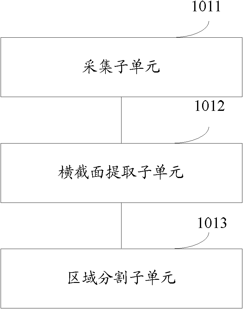
[0046] A region extraction unit 102, configured to extract the region of the blood vessel to be detected from the blood vessel image of the enhanced coronary computed tomography image CTA;

[0047] A morphological feature extraction unit 103, configured to extract the same...

Embodiment 2

[0132] The embodiment of the present application also provides a detection device for a deformed area of ​​a blood vessel, which is different from the first embodiment above in that the embodiment of the present application further includes a morphological feature output unit. see Figure 14 , which is a structural schematic diagram of another embodiment of a device for detecting a deformed region of a blood vessel in the present application. In addition to including a classifier construction unit 101, a region extraction unit 102, a morphological feature extraction unit 103 and a detection unit 104, the device In addition, it also includes a morphological feature output unit 105, wherein,

[0133] The classifier construction unit 101 is configured to use the morphological features reflecting the boundary shape and / or area shape of the blood vessel region as the classification basis, and establish a classifier according to the data classification model, wherein the feature spa...

Embodiment 3

[0143] Corresponding to the above-mentioned detection device for the deformation region of blood vessels, the embodiment of the present application also provides a detection method for deformation of blood vessels, please refer to Figure 15 , which is a flowchart of an embodiment of a method for detecting a deformed region of a blood vessel in the present application, the method includes the following steps:

[0144] Step 1501: Taking the morphological features reflecting the boundary shape and / or regional shape of the blood vessel region as the classification basis, and establishing a classifier according to the data classification model in advance;

[0145] The constructed classifier can classify the vessel region into two types with deformation or without deformation according to various morphological features, so as to realize the detection function of the blood vessel to be detected. As we all know, data classification is an important content in data mining. Common data ...


PUM


Abstract

The embodiment of the invention discloses a device and a method for detecting a blood vessel deformation area. The device comprises a classifier constructing unit, an area extracting unit, a morphological characteristic extracting unit and a detecting unit, wherein the classifier constructing unit is used for establishing a classifier according to a data classification model by taking morphological characteristics reflecting a border shape and / or an area shape in a blood vessel area as a classification basis; a characteristic space in the classifier is used for marking the presence or absence of deformation of the blood vessel area; the area extracting unit is used for extracting a blood vessel area to be detected from a blood vessel image of a computed tomography angiography (CTA); the morphological characteristic extracting unit is used for extracting morphological characteristics same as the morphological characteristics in the classifier in the blood vessel area to be detected; and the detecting unit is used for identifying the characteristic space corresponding to the extracted morphological characteristics in the classifier, and acquiring a deformation detection result of the blood vessel area to be detected according to the corresponding characteristic space. The device and the method can improve the sensitivity and the accuracy of the detection result.

Description

technical field [0001] The present application relates to medical image technology, in particular to a detection device and method for a deformed area of ​​a blood vessel. Background technique [0002] In the field of medical images, CTA (Computed Tomography Angiography) has become the main screening method for analyzing the internal morphology of the human body due to its high temporal and spatial resolution and non-invasiveness. By processing the CTA, it is convenient for people to quickly find an area of ​​interest. For example, detection of vessel deformation regions in CTA vessel images. [0003] At present, among various medical devices for detecting the deformed area of ​​blood vessels in CTA blood vessel images, a widely used method is: through computer technology and image processing technology, computer-aided measurement of the deformed position of the vessel lumen in CTA blood vessel images and degree of deformation. The detection method further includes diamet...

Claims


Application Information

Patent Timeline
IPC IPC(8): G06K9/62
Inventor 王胜军赵凯黄艳康雁
Owner SHENYANG NEUSOFT MEDICAL SYST CO LTD
Why Eureka
Try Eureka