
Ply tissue engineering corneal frame and manufacturing method and application thereof

A tissue engineering, lamellar cornea technology, used in medical science, eye implants, prostheses, etc., can solve biocompatibility, strength, degradation rate, transparency, homology, antigenicity and pathogenicity Defects and other problems, to achieve the effect of simple and reliable production process, reduced damage to the matrix, and a wide range of sources

Active Publication Date: 2011-01-19
XIAMEN DAKAI BIOTECH CO LTD
View PDF4 Cites 28 Cited by
  • Summary
  • Abstract
  • Description
  • Claims
  • Application Information

AI Technical Summary

Problems solved by technology

However, various seed cell scaffold materials developed at home and abroad have some defects in corneal curvature and topography, biocompatibility, strength, degradation rate, transparency, homology, antigenicity and pathogenicity.

Method used


Image

  • Ply tissue engineering corneal frame and manufacturing method and application thereof
  • Ply tissue engineering corneal frame and manufacturing method and application thereof
  • Ply tissue engineering corneal frame and manufacturing method and application thereof

Examples


Embodiment 1

[0046] The lamellar tissue engineering corneal stent described in the present invention, taking the dry and rehydrated lamellar corneal stent as an example, comprises the following steps: after the fresh animal eyeball is disinfected with iodine, it is treated with alcohol and scraped to remove the epithelial cells, and the Lamellar cornea, remove the lamellar corneal stroma, put it on the corneal pillow and use a corneal trephine to remove the lamellar corneal slices of different diameters, through repeated regular airtight freeze-thaw, hypotonic swelling, DNA, RNase digestion, airtight Ice bath electrophoresis in a sterilization box, drying technology in a 24-well plate, cobalt 60 disinfection technology, etc., and finally the lamellar corneal stroma sheet that was not dried, dried, and rehydrated after drying was obtained to obtain the lamellar tissue engineered corneal stroma scaffold. Save it for later use, and add the required growth factors before use.

[0047] Preparat...

Embodiment 2

[0050] The lamellar tissue engineered corneal stent described in the present invention, taking the dry and rehydrated lamellar corneal stent as an example, includes the following steps: after the fresh animal eyeball is sterilized with antibiotics, it is treated with alcohol and scraped to remove the epithelial cells, and the lamellar tissue is separated. Cornea, remove the lamellar corneal stroma sheet, put it on the corneal pillow and use the corneal trephine to remove the lamellar corneal sheet of different diameters, through repeated regular sealing freeze-thawing, hypotonic swelling, DNA, RNase digestion, airtight disinfection In-box ice-bath electrophoresis, 24-well plate drying technology, cobalt 60 disinfection technology, etc., finally obtained undried, dried and rehydrated lamellar corneal stroma sheets to obtain the lamellar tissue engineered corneal stroma scaffold. Save it for later use, and add the required growth factors before use.

[0051]The first preparation...


PUM

PropertyMeasurementUnit
Tensile strengthaaaaaaaaaa
Depthaaaaaaaaaa
Thicknessaaaaaaaaaa

Abstract

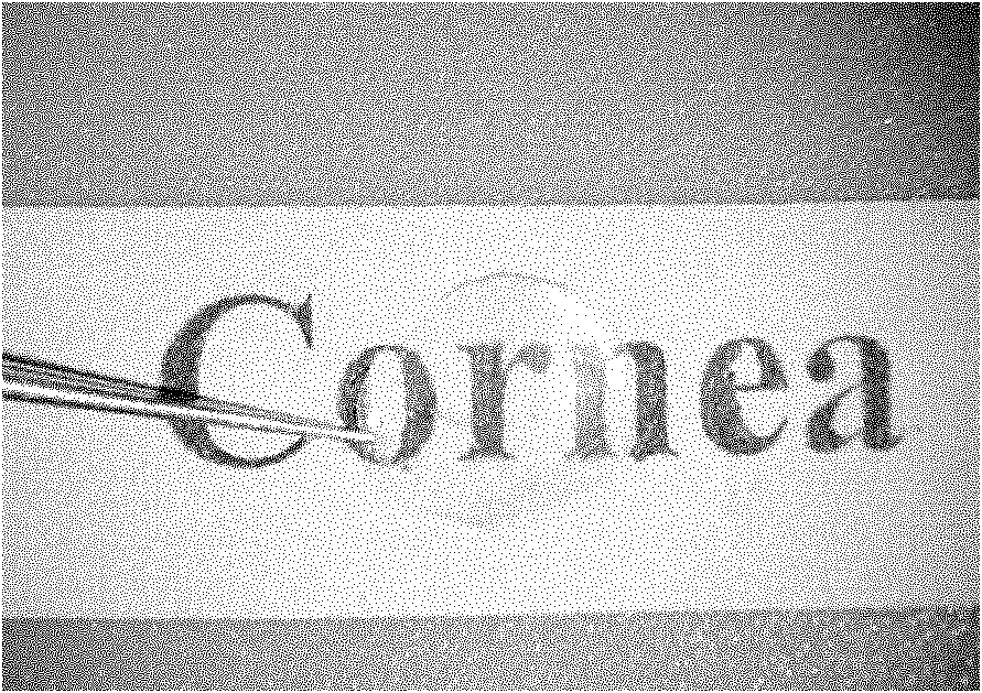
The invention discloses a ply tissue engineering corneal frame and a manufacturing method and an application thereof, relating to a ply tissue engineering corneal frame of the biomedicine. Compared with the existing materials, the invention provides a ply tissue engineering corneal frame and a manufacturing method and an application thereof, which has abundant resources, high transparency, good biocompatibility, thorough decellularization and strong safety, and the performance approaches to that of a fresh cornea, so that the ply tissue engineering corneal frame can be accepted by majority of patients and applied clinically for a long term. The ply tissue engineering corneal frame is an animal derived decellularized ply corneal stroma sheet and does not contain cellular constituents; collagenous fibers are tidily arranged, and gaps are regular; corneal light transmittance is 80-95%, and tensile strength is 2-5N / mm<2>. The ply tissue engineering corneal frame can be served as the substitute for various donor materials for corneal transplantation and can be used for treating a series of diseases of corneal trauma and chemical burn, corneal tumor and a series of hyperplastic diseases, a series of diseases of corneal neovascularisation and scar, corneal immune diseases, a series of diseases caused by corneal transplantation exclusive reaction and other keratopathy.

Description

technical field [0001] The invention relates to a biomedical lamellar tissue engineering corneal scaffold, in particular to a scaffold for treating corneal trauma, tumors, chemical burns (alkali burns), vascularized severe dry eye, limbal stem cell deficiency, and immune diseases Such as Stevens-Johnson syndrome (SJS), ocular pemphigoid, herpes simplex virus leukoplakia, corneal transplant rejection and other blinding eye diseases such as lamellar corneal tissue engineering materials and their preparation methods and applications. Background technique [0002] According to the report of the World Health Organization, corneal disease is the second major cause of vision loss. Every year, 1.5 million to 2 million new corneal blindness are caused by corneal ulcers and eye trauma. The main cause of childhood blindness is corneal blindness caused by measles. Vitiligo. The only effective treatment for corneal blindness is keratoplasty. However, due to the influence of religious b...

Claims


Application Information

Patent Timeline
IPC IPC(8): A61F2/14A61L27/38
Inventor 刘祖国李炜邵毅许传超梁凌毅
Owner XIAMEN DAKAI BIOTECH CO LTD
Learn More