Eureka AIR delivers breakthrough ideas for toughest innovation challenges, trusted by R&D personnel around the world.

PHBV (Poly (HydroxyButyrate-hydroxyValerate)) nano fiber support material as well as preparation method and application thereof

A scaffold material and nanofiber technology, applied in medical science, prosthesis, etc., can solve problems such as lack of mechanical properties of 3D structures, few successful examples of renewable scaffolds, lack of geometric complexity, etc., to achieve scaffold structure and fiber morphology Controllable, inexpensive, adhesion-promoting effect

Active Publication Date: 2010-12-29
SOUTH CHINA UNIV OF TECH
View PDF3 Cites 12 Cited by
  • Summary
  • Abstract
  • Description
  • Claims
  • Application Information

AI Technical Summary

Problems solved by technology

Self-assembly is generally hydrogel, which lacks geometric complexity and lacks mechanical properties in 3D structures, so there are few successful examples in the construction of 3D complex regenerative scaffolds
There is no report on the preparation of PHBV nanofibers by thermally induced phase separation.

Method used


Image

  • PHBV (Poly (HydroxyButyrate-hydroxyValerate)) nano fiber support material as well as preparation method and application thereof
  • PHBV (Poly (HydroxyButyrate-hydroxyValerate)) nano fiber support material as well as preparation method and application thereof
  • PHBV (Poly (HydroxyButyrate-hydroxyValerate)) nano fiber support material as well as preparation method and application thereof

Examples


Embodiment 1

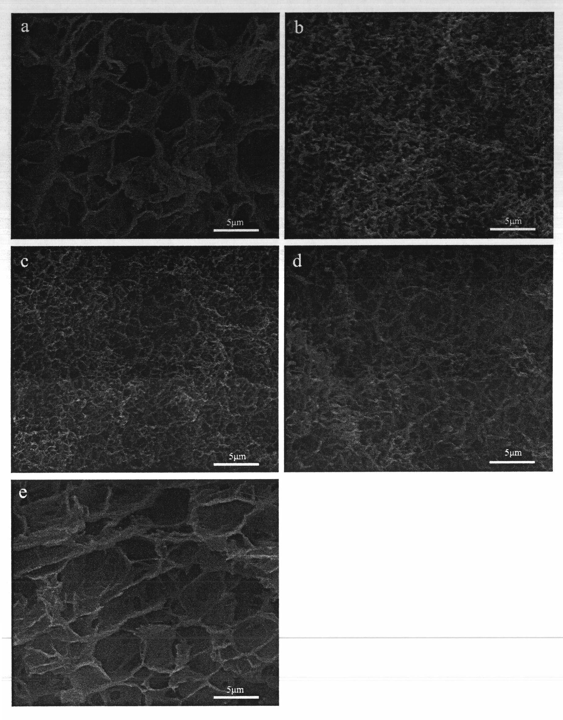
[0028] Chloroform: dioxane (9:1) solution to prepare PHBV pore-wall scaffolds ( figure 1 a)

[0029] Take the molecular weight as 3×10 5 Dissolve 0.225g of PHBV powder in 2.7ml chloroform solution, stir magnetically at 50°C for 3 hours, add 0.3ml dioxane solution, continue stirring for 2 hours, transfer to a 5ml beaker, seal with sealing glue, and place at -24°C Freeze in the refrigerator for 12 hours. Add acetone solution to replace the organic solvent, place in a 4°C refrigerator, change the acetone twice a day for 3 days; use deionized water to replace the acetone solution, place in a 4°C refrigerator, change the water twice a day, and replace for 3 days. Samples were freeze-dried at -80°C for 24 hours.

[0030] SEM image of PHBV scaffold ( figure 1 a) shows that it has a pore-wall structure, the thickness of the pore wall is 150-300 nm, and the pore diameter is 1.0-6.5 μm.

Embodiment 2

[0032] The PHBV sheet structure scaffold was prepared in chloroform:dioxane (8:2) solution ( figure 1 b)

[0033] Take the molecular weight as 3×10 5 Dissolve 0.225g of PHBV powder in 2.4ml chloroform solution, stir magnetically at 50°C for 3 hours, add 0.6ml dioxane solution, continue stirring for 2 hours, transfer to a 5ml beaker, seal with sealing glue, and place at -24°C Freeze in the refrigerator for 12 hours. Add acetone solution to replace the organic solvent, place in a 4°C refrigerator, change the acetone twice a day for 3 days; use deionized water to replace the acetone solution, place in a 4°C refrigerator, change the water twice a day, and replace for 3 days. Samples were freeze-dried at -80°C for 24 hours.

[0034] SEM image of PHBV scaffold ( figure 1 b) shows that it has a lamellar structure with a thickness of 60-120 nm and a pore size of 0.5-1.5 μm.

Embodiment 3

[0036] PHBV nanofibrous scaffolds were prepared in chloroform:dioxane (7:3) solution ( figure 1 c)

[0037] Take the molecular weight as 3×10 5 Dissolve 0.225g of PHBV powder in 2.1ml of chloroform solution, stir magnetically at 50°C for 3 hours, add 0.9ml of dioxane solution, continue stirring for 2 hours, transfer to a 5ml beaker, seal with sealing glue, and place at -24°C Freeze in the refrigerator for 12 hours. Add acetone solution to replace the organic solvent, place in a 4°C refrigerator, change the acetone twice a day for 3 days; use deionized water to replace the acetone solution, place in a 4°C refrigerator, change the water twice a day, and replace for 3 days. Samples were freeze-dried at -80°C for 24 hours.

[0038] SEM image of PHBV scaffold ( figure 1 c) shows that it is a nanofiber structure, the diameter of the nanofiber is 40-100 nm, and the pore size is 0.3-1.0 μm.


PUM

PropertyMeasurementUnit
Thicknessaaaaaaaaaa
Diameteraaaaaaaaaa
Aperture sizeaaaaaaaaaa

Abstract

The invention discloses a PHBV (Poly (HydroxyButyrate-hydroxyValerate)) nano fiber support material, a preparation method and application thereof. The preparation method comprises the following steps of: (1) dissolving PHBV powder with the molecular weight of 3*105 in an organic solvent to compound a solution with the concentration of 7.5%, and then stirring for 3-5 hours in a water bath at 50 DEG C by using magnetic force; (2) after fully dissolving the PHBV, hermetically placing the solution; (3) after the solution fully forms hydrogel, taking the hydrogel out, adding acetone for replacing the organic solvent, and then replacing the acetone by using deionized water; and (4) and freezing and drying the PHBV hydrogel to obtain the PHBV nano fiber support material. The PHBV nano fiber support material has the advantages of low cost, simple preparation process, good biocompatibility, high porosity and controllable support structure and fiber morphology; and because the morphological structure of the PHBV nano fiber support material is similar to that of an extracellular matrix in cartilaginous tissue cells of a human body, the PHBV nano fiber support material can promote the adhesion, the propagation and the differentiation of cartilaginous cells and is very suitable for restoring and reconstructing cartilaginous tissues.

Description

technical field [0001] The invention belongs to the technical field of nanofiber preparation, and in particular relates to the thermally induced phase separation technology of polymer solutions. Background technique [0002] Cartilage itself does not contain blood vessels, lymph, and lacks the ability to spontaneously regenerate. The current treatment methods for cartilage damage have certain limitations. Therefore, in 1987, tissue engineering was formally proposed as a new discipline. The basic technical route of cartilage tissue engineering is to culture seed cells in vitro and plant them on a suitable scaffold with good biocompatibility and degradability to form a cell-scaffold complex. Implanted into the tissue defect site in the living body, and finally complete the repair and reconstruction of the tissue. [0003] Degradable biomaterials play an important role in tissue engineering research, as temporary 3D scaffolds play a guiding role in the process of tissue repai...

Claims


Application Information

Patent Timeline
IPC IPC(8): A61L27/18A61L27/50
Inventor 王迎军陈聪杜昶
Owner SOUTH CHINA UNIV OF TECH
Why Eureka
Eureka Blog
Learn More