Colorimetric detection method based on nanometer-gold and nucleic acid structure and kit thereof
A detection method, nano-gold technology, which is applied in the direction of material analysis by observing the influence of chemical indicators, and analysis by making materials undergo chemical reactions, can solve problems such as inapplicability, and achieve low cost, high specificity, The effect of improving sensitivity
- Summary
- Abstract
- Description
- Claims
- Application Information
AI Technical Summary
Problems solved by technology
Method used
Image
Examples
Embodiment 1
[0040] The detection of embodiment 1 cocaine
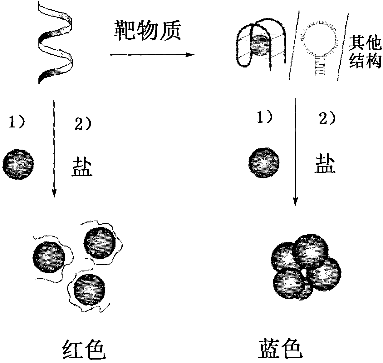
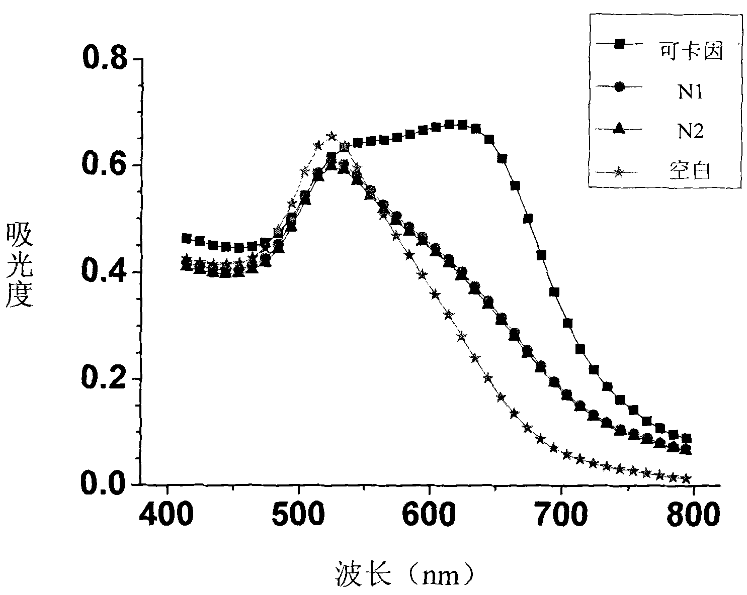
[0041] Steps: Take 2 μL of cocaine-nucleic acid aptamer solution with a concentration of 100 μM and 2 μL of an aqueous solution of cocaine hydrochloride with a concentration of 0.1 to 10 mM, add 18 μL of buffer solution (25 mM Tris, pH 8.2, 0.6 M NaCl) and mix ( The final concentration of DNA is 10 μM, the final concentration of cocaine is 10-1000 μM), and the reaction is carried out at room temperature for 30 min. At the same time, the group without adding cocaine and adding 2 μL of water was used as a blank, and the two groups of adding 2 μL of 1 mM benzoylecgonine (N1) and ecgonine methyl ester (N2) were used as controls. Then take 2 μL of the above reaction solution and add 100 μL of the nano-gold solution (13nm, 3.5nM) prepared above. ) (final concentration about 50mM). Observe the color change of gold nanoparticles in the experimental group and the control group and record the ultraviolet-visible spectrum.
[0042] Result...
Embodiment 2
[0043] The detection of embodiment 2ATP
[0044] Steps: Take 2 μL of ATP-nucleic acid aptamer solution with a concentration of 10 μM and 2 μL of an aqueous solution of ATP with a concentration of 0.01 to 1 mM, add 18 μL of buffer solution (10 mM Tris, pH 8.2, 0.3 M NaCl) and mix (final DNA concentration 1 μM, the final concentration of ATP is 1-100 μM), and reacted at room temperature for 15 minutes. At the same time, a group without adding ATP and adding 2 μL of water was used as a blank, and a group of adding 2 μL of 1 mM CTP, UTP, and GTP was used as a control. Then take 2 μL of the above-mentioned reaction solution and add 10 μL of the nano-gold solution (20nm, 2nM) prepared above, and react at room temperature for 5 minutes, then add 10 μL of 0.2M PBS (10mM PB, pH7.0, 0.2M NaCl) (PBS The final concentration is approximately 100 mM). The color changes and ultraviolet-visible spectra of the gold nanoparticles in the experimental group and the control group were recorded r...
Embodiment 3
[0046] Embodiment 3 is to the detection of divalent mercury ion
[0047] Step: Take 2 μL of Hg with a concentration of 100 μM 2+ - MSO solution, add 2 μL of Hg with a concentration of 0.1-2 mM 2+ aqueous solution (the reaction concentration of MSO is 50 μM, the reaction concentration of the target substance is 50-1000 μM, the reaction buffer solution and the ionic strength are both 0), react at room temperature for 1 min. without adding Hg 2+ , add 2 μL of water to a group as a blank experiment. In addition, selectivity analysis was carried out through two groups of experiments, one group added 2 μL of 25 mM Ca2+ , Mg 2+ The other group added 0.5mM mixed ions (Fe 2+ , Cu 2+ ,Co 2+ , Mn 2+ , Ni 2+ , Zn 2+ , Cd 2+ ). Then take 2 μL of the above reaction solution and add 100 μL of the nano-gold solution (10nm, 100nM) prepared above, and react at 37°C for 1min, then add 60μL of 1M NaNO 3 (Final concentration of salt is approximately 600 mM). Observe the color change o...
PUM
| Property | Measurement | Unit |
|---|---|---|
| particle diameter | aaaaa | aaaaa |
Abstract
Description
Claims
Application Information
 - Generate Ideas
- Intellectual Property
- Life Sciences
- Materials
- Tech Scout
- Unparalleled Data Quality
- Higher Quality Content
- 60% Fewer Hallucinations
Browse by: Latest US Patents, China's latest patents, Technical Efficacy Thesaurus, Application Domain, Technology Topic, Popular Technical Reports.
© 2025 PatSnap. All rights reserved.Legal|Privacy policy|Modern Slavery Act Transparency Statement|Sitemap|About US| Contact US: help@patsnap.com


