Medical ultrasonic image line detection method
A technology for ultrasonic image and line detection, which is applied in the field of medical ultrasonic image line detection, and can solve the problems of large influence of subjective factors on detection results, poor anti-noise performance, poor detection effect of low signal-to-noise ratio images, etc.
- Summary
- Abstract
- Description
- Claims
- Application Information
AI Technical Summary
Problems solved by technology
Method used
Image
Examples
Embodiment 1
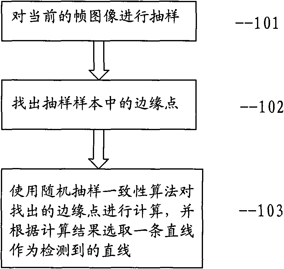
[0035] The medical ultrasound image straight line detection method of the present invention, such as figure 1 shown, including the following steps:
[0036] 101. Sampling the current frame image;
[0037] Wherein, the sampling process is as follows: the current frame image is vertically sampled according to a preset sampling interval. A positive integer N is preset as the sampling interval, and then a single column of pixels is selected vertically every N pixels in the horizontal direction as a sampling sample. Line detection will be achieved through these sampling samples.
[0038] 102. Find the edge point in the sampling sample; that is, set a grayscale threshold T1, and select the first pixel whose grayscale value exceeds the grayscale threshold T1 from top to bottom after sampling the sample difference as an edge point , if none of them exceeds the gray threshold T1, the point with the maximum gray value is selected as the edge point.
[0039] 103. Use the random sampl...
Embodiment 2
[0042] The medical ultrasound image straight line detection method of the present invention, such as figure 2 shown, including the following steps:
[0043] 201. Set an area of interest;
[0044] That is: before starting real-time detection, form a convex polygon by selecting the vertices of the polygon in the image displayed on the screen (circle or ellipse belongs to a special case of convex polygon, so the convex polygon in the present invention includes circle or ellipse) , the region within the convex polygon is the region of interest, and the subsequent line detection step will only be performed on the image within the region of interest. (Such as Figure 4 )
[0045] 202. Sampling the region of interest in the current frame image;
[0046] The sampling process is as follows: the current frame image is vertically sampled according to the preset sampling interval. Preset a positive integer N as the sampling interval, and then vertically select a single column of p...
Embodiment 3
[0051] The medical ultrasound image straight line detection method of the present invention, such as image 3 shown, including the following steps:
[0052] 301. Set an area of interest;
[0053] That is: before starting real-time monitoring, a convex polygon is determined by selecting the vertices of the convex polygon in the image displayed on the screen, and the area inside the convex polygon is the area of interest, and the subsequent line detection steps will only be performed on the image in the area of interest. . (Such as Figure 4 ).
[0054] 302. Sampling the region of interest in the current frame image;
[0055] The sampling process is as follows: the current frame image is vertically sampled according to the preset sampling interval. Preset a positive integer N as the sampling interval, and then vertically select a single row of pixels in the region of interest every N pixels in the horizontal direction as sampling samples. Line detection will be achie...
PUM
 Abstract
Description
Claims
Application Information
 - R&D
- Intellectual Property
- Life Sciences
- Materials
- Tech Scout
- Unparalleled Data Quality
- Higher Quality Content
- 60% Fewer Hallucinations
Browse by: Latest US Patents, China's latest patents, Technical Efficacy Thesaurus, Application Domain, Technology Topic, Popular Technical Reports.
© 2025 PatSnap. All rights reserved.Legal|Privacy policy|Modern Slavery Act Transparency Statement|Sitemap|About US| Contact US: help@patsnap.com


