
Target cell staining kit for displaying proliferative activity of cell and use method thereof

A technology targeting cells and dyeing reagents, which is applied in biochemical equipment and methods, and microbial measurement/inspection, etc., can solve the problems of affecting the observation of enzyme reaction products, high cost of kit material packaging, and narrow application range, etc., to achieve improvement Staining effect and film reading effect, saving time of azo process, effect of reducing cell layer thickness

Active Publication Date: 2010-07-21
泰普生物科学(中国)有限公司
View PDF0 Cites 11 Cited by
  • Summary
  • Abstract
  • Description
  • Claims
  • Application Information

AI Technical Summary

Problems solved by technology

[0004] Yet this method of operation has the following disadvantages in application: 1, the preparation process of the enzyme incubation solution is numerous and diverse, and shows that (1) azo salts will be temporarily prepared with sodium nitrite, which needs to be prepared in the 46ml and 2.5ml acetic acid containing Preheating the reaction in the flask for 3 minutes is time-consuming and laborious
(2) 10 drops of reagents need to be added each time to prepare various reagents, which is prone to errors in actual use
2. The incubation time is longer, it takes 45 minutes
3. Papanicolaou staining is required for counterstaining, which increases the operation steps and affects the observation of enzyme reaction products
4. The whole dyeing time is too long, the whole process takes more than 60 minutes
5. The scope of application is narrow, only for cervical cells
5. The cost of kit materials and packaging is relatively high

Method used


Image

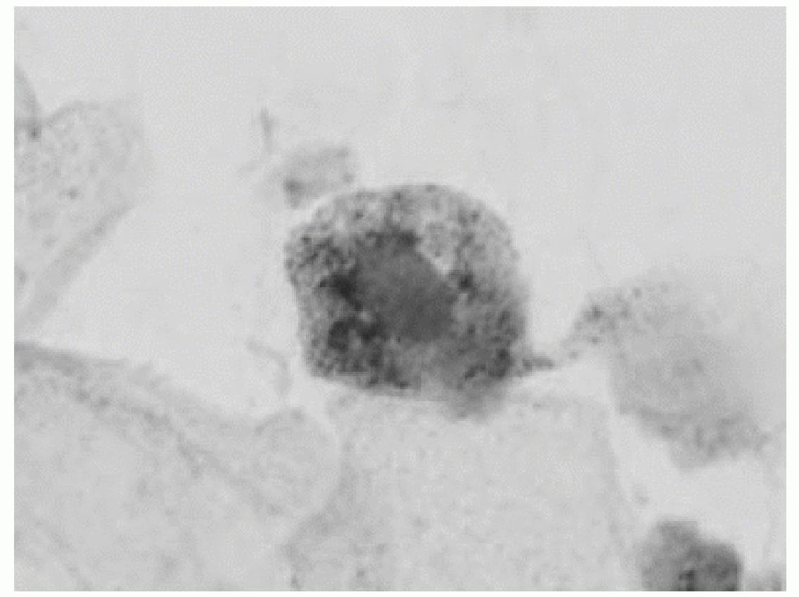
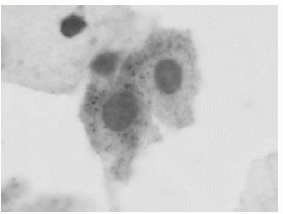
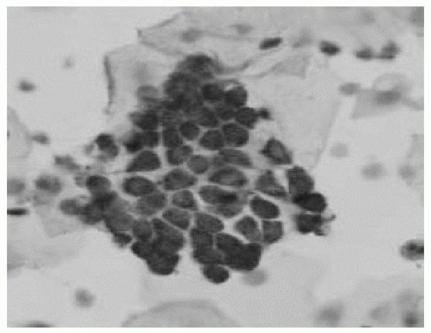
  • Target cell staining kit for displaying proliferative activity of cell and use method thereof
  • Target cell staining kit for displaying proliferative activity of cell and use method thereof
  • Target cell staining kit for displaying proliferative activity of cell and use method thereof

Examples


Embodiment 1

[0113] Example 1: Cervical Cancer Screening

[0114] Test design

[0115] 1. Experimental group: Cervical exfoliated cell smear materials come from daily outpatient gynecological cases and scattered gynecological examination population in the physical examination department. Use Fujian Taipu Bioscience Co., Ltd. preservation solution and sampling brush to sample in the cervical transition zone according to the standard, and perform staining and preparation according to the method of the present invention (details will be introduced later).

[0116] 2. Control group: use the same specimen to prepare another smear (that is, prepare two cell smears at the same time from one specimen, and divide the cells equally when making the smear), and use conventional Pap staining.

[0117] 3. Film reading and registration: The double-blind method was used to read the film. The experimental group was observed by the physicians of the research group. The diagnosis was based on TBS terms. The...

Embodiment 2

[0148] Embodiment 2 retrospective statistical data

[0149] From the computer system of the cervical cytology screening data of the Pathology Department of Xiamen Second Hospital, the diagnostic results of using TBS diagnostic terms after adopting the staining solution of the present invention on September 1, 2009-11.26, and using the same period in 2008 The results (see Table 5) showed that targeted staining had a significant effect on atypical squamous cells, atypical glandular cells, low-grade intraepithelial lesions, high-grade intraepithelial lesions and the detection rate of HPV (26.8 %) was significantly higher than traditional Pap staining (11.81%).

[0150] Table 5 The detection rate of targeted staining in 2009 and Pap staining in 2008

[0151]

Embodiment 3

[0152] Embodiment 3, esophageal brushing to get exfoliated cells

[0153] 39 cases of progressive targeted staining (staining method is the same as Example 1) of the esophageal brush cell smears submitted to the endoscopy room of the Second Hospital of Xiamen City, and compared with conventional Pap staining, the results (Table 6.) It shows that the positive detection rate of the experimental group is higher than that of the Papanicolaou group.

[0154] Table 6 Comparison of targeted staining and traditional Pap staining results in 39 cases of esophageal brush slides

[0155]


PUM

PropertyMeasurementUnit
Sensitivityaaaaaaaaaa

Abstract

The invention discloses a target cell staining kit for displaying the proliferative activity of a cell and a use method thereof. The kit comprises I type and II type which respectively comprise an enzyme substrate, test paper, enzyme stationary liquid, phosphate buffer solution, Mayer hematoxylin and the like. The staining method comprises the steps of preparing pieces, fixing, staining and the like. The method can display acid phosphatase in exfoliative cells on smears such as exfoliative cervical cells, esophagus brush sheet cells and the like and search abnormal proliferated cells according to the target. Compared with the traditional Pasteur smear method, the method has the advantages of higher sensitivity for detecting cancer and precancerous lesions and similar specificity, therefore, the invention can be used for cytology screening of cervical carcinoma, esophageal cancer, lung cancer and other malignant tumors.

Description

technical field [0001] The present invention relates to a kit for displaying cell proliferation activity and its use method, in particular to a method and kit for displaying cell acid phosphatase on smears of exfoliated cells such as cervical exfoliated cells and esophageal brush cells. Background technique [0002] Since the middle of the last century, Pap smear and staining techniques have been gradually promoted as a screening method for cervical cancer. Significant reductions in cervical cancer mortality and incidence have been observed in countries where this approach has been applied. Of the more than 50 million routine Pap tests performed in the United States each year, 3.5 million (7%) are positive / abnormal. However, there are still 4,500 deaths and 13,000 new cases of cervical cancer each year1. Studies have shown that 20% of new cancer cases in the United States have never been tested positive by Pap stain, and there are 3 to 5 years before the onset of disease. ...

Claims


Application Information

Patent Timeline
Patent Type & Authority Applications(China)
IPC IPC(8): C12Q1/42
Inventor 谢佐福
Owner 泰普生物科学(中国)有限公司
Learn More