
Application of amniotic epithelial cells in pharmacy

A technology of amniotic membrane epithelial cells and uses, applied in the field of biomedicine, can solve problems such as limited clinical application and source problems, and achieve the effects of wide sources, improvement of mobility function, and reduction of cerebral edema symptoms

Active Publication Date: 2010-06-09
SHANGHAI ICELL BIOTECH +5
View PDF6 Cites 4 Cited by
  • Summary
  • Abstract
  • Description
  • Claims
  • Application Information

AI Technical Summary

Problems solved by technology

The use of stem cells to repair or regenerate damaged brain nerve cells has become a hot spot in the field of neuroscience today. The transplantation of neural stem cells and embryonic stem cells once opened up a new way for the treatment of brain injuries, but due to the source of the two, the clinical application is limited.

Method used


Image

  • Application of amniotic epithelial cells in pharmacy
  • Application of amniotic epithelial cells in pharmacy
  • Application of amniotic epithelial cells in pharmacy

Examples


Embodiment 1

[0042] Example 1. hAEC isolation and culture

[0043] Take the placenta of healthy caesarean section pregnant women, and strip the amniotic membrane from the chorion. Rinse with PBS to remove blood cells, digest with 0.25% trypsin at 37°C for 30 min, pipette, add RPMI1640 containing 10% FBS to stop the digestion, filter through a 100-mesh sieve, centrifuge at 1,500 rpm, discard the supernatant, and dilute the mixture with 1×10 4 / cm 2 Inoculate in 10cm dish. hAECs were placed in RPMI-1640 (10% FBS, 100 μg / ml streptomycin, 100 U / ml penicillin, 10 ng / ml EGF and 0.3 mg / ml glutamine) at 37°C, 5% CO 2 cultured in a cell culture incubator.

Embodiment 2

[0044] Example 2. Preparation of hAEC cell preparation

[0045] Example 1 hAEC cultured for 24-72 hours, remove the medium, digest the adherent hAEC with 0.25% trypsin and centrifuge, then rinse with 2ml of PBS solution and centrifuge again, resuspend in PBS and adjust the cell concentration to 105 / μl for later use, namely hAECs cell preparations were obtained.

Embodiment 3

[0046] Example 3. hAECs treat cerebral hemorrhage model in rats

[0047] 1 animal

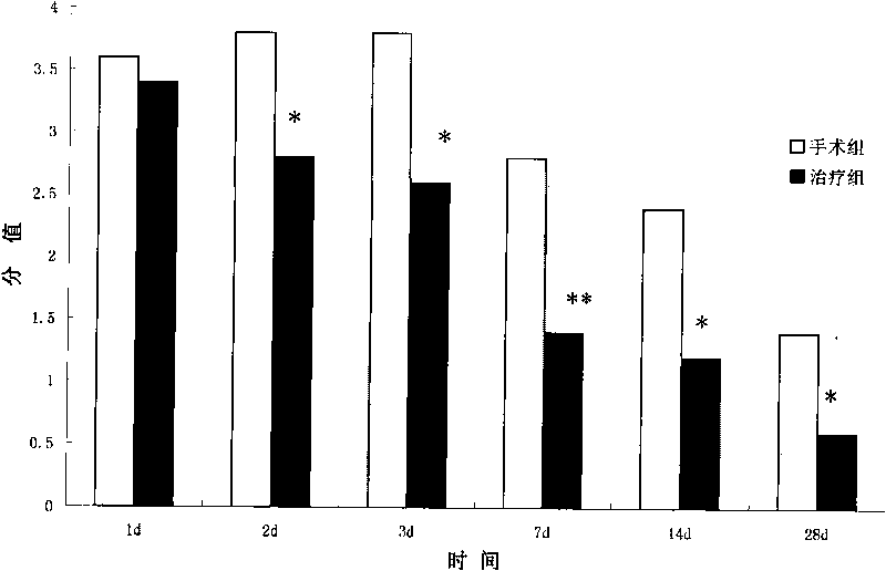
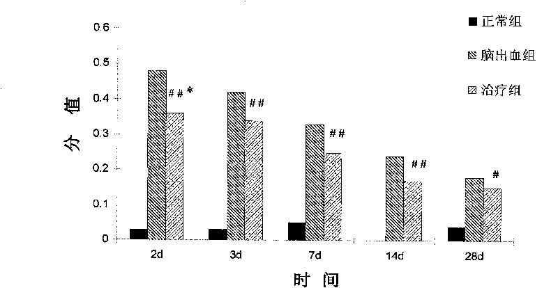
[0048] SPF grade female adult Sprague-Dawley rats were used in the experiment, purchased from Shanghai Slack Experimental Animal Center, weighing 250±10g, reared in a single cage, free to ingest water and food, and lighted to form a day / night cycle of 12h-12h. The experimental animals were divided into control group, operation (cerebral hemorrhage) group and treatment (cell transplantation) group, wherein the operation group and treatment group were divided into post operation and treatment 2d, 3d, 7d, 14d and 28d.

[0049] 2 Research Methods

[0050] (1) Preparation of cerebral hemorrhage model in rats

[0051] Refer to the methods of CrystalL [Crystal L MacLellan, Gergely Silasi, Candice C Poon, et al. Journal of Cerebral Blood Flow & Metabolism, 2007: 1-10] and make improvements. Rats were anesthetized with chloral hydrate (10% chloral hydrate, injected intraperitoneally by 0.3ml / 100g bod...


PUM


Abstract

The invention relates to the technical field of biomedicine, in particular to application of amniotic epithelial cells in a medicament for treating hemorrhagic cerebral vascular diseases. The invention discloses the application of the amniotic epithelial cells in treating or improving the hemorrhagic cerebral vascular diseases. The amniotic epithelial cells can effectively treat or improve the hemorrhagic cerebral vascular diseases, obviously improve the action function of patients with the hemorrhagic cerebral vascular diseases, and obviously reduce encephaledema symptoms caused by the hemorrhagic cerebral vascular diseases; the treatment do not need match because the amniotic epithelial cells do not express or weakly express MHC; the amniotic epithelial cells also do not express telomerase, and cell proliferation is controlled, so that the transplantation treatment is safe; meanwhile, the amniotic epithelial cells have wide sources, so that the phenomenon of ethic or morality is avoided. Therefore, the amniotic epithelial cells provide a new effective choice for clinically treating or improving the hemorrhagic cerebral vascular diseases in the future.

Description

technical field [0001] The invention relates to the technical field of biomedicine, in particular to the use of amniotic epithelial cells in medicine for treating hemorrhagic cerebrovascular diseases. Background technique [0002] Cerebrovascular disease refers to a group of diseases with cerebral hemorrhagic or ischemic injury symptoms as the main clinical manifestation caused by cerebral blood vessel rupture, bleeding or thrombosis, also known as cerebrovascular accident or stroke, commonly known as cerebral apoplexy. The disease is often seen in the acute attack of middle-aged people, and severe cases may cause disturbance of consciousness and limb paralysis. Cerebrovascular disease can be divided into two categories according to its nature. One is ischemic cerebrovascular disease, which is more common in clinical practice and accounts for about 70% to 80% of all cerebrovascular patients. It is caused by cerebral arteriosclerosis and other reasons. Cerebral artery lumen ...

Claims


Application Information

Patent Timeline
IPC IPC(8): A61K35/50A61P9/10
Inventor 郭礼和刘新洪燕龙尚婧瑨庞力
Owner SHANGHAI ICELL BIOTECH
Learn More