
Artificial uterus three-dimensional gel model used for researching development and differentiation of embryonic stem cells

A technology of embryonic stem cells and uterus, applied in a series of key technical fields, can solve problems such as lack of bionic conditions

Inactive Publication Date: 2010-05-26
INST OF BASIC MEDICAL SCI ACAD OF MILITARY MEDICAL SCI OF PLA
View PDF0 Cites 7 Cited by
  • Summary
  • Abstract
  • Description
  • Claims
  • Application Information

AI Technical Summary

Problems solved by technology

Intersecting with the traditional two-dimensional differentiation and development culture, the three-dimensional model provides an effective development space for further research on the differentiation and development of embryonic stem cells, but the simple three-dimensional scaffold is still very different from the differentiation and development environment of the embryonic inner cell mass in the uterus. None of these models are well suited for biomimicry

Method used


Image

  • Artificial uterus three-dimensional gel model used for researching development and differentiation of embryonic stem cells
  • Artificial uterus three-dimensional gel model used for researching development and differentiation of embryonic stem cells
  • Artificial uterus three-dimensional gel model used for researching development and differentiation of embryonic stem cells

Examples


Embodiment 1

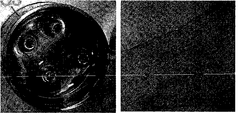
[0041] The workflow of the three-layer tissue-engineered artificial uterine sheet constructed in vitro is as follows: figure 1 shown. The details are as follows: Mix the type I liquid collagen and Matrigel glue evenly at a ratio of 4:1, then mix it with the DMEM-F12 concentrated culture solution containing uterine smooth muscle cells in equal volume, and adjust the pH value of the mixture to 0.1M NaOH. 7.2 to 7.4, add to the prepared 12-hole plate mold, inject 0.4ml into each hole. Place it at 37°C, 5% CO 2 After incubating in the incubator with 5mim liquid complex solidified, take it out. According to the same method, uterine stromal cells were added on the surface of the solidified smooth muscle cells, and after solidification, endometrial epithelial cells were added in the same method. Make a three-dimensional three-dimensional artificial uterus three-dimensional gel model (such as figure 2 shown). Add estrogen and progesterone to regulate the cycle of uterine prolife...

Embodiment 2

[0042] Example 2. Embryonic stem cells were introduced into the in vitro bionic model in two ways: surface inoculation or internal uniform compounding:

[0043] 1) Inoculate embryonic stem cells on the surface of uterine slices: inoculate embryonic stem cells in the form of single cells on the surface of prepared uterine slices (such as image 3 shown), to observe the embryoid body-like embryoid stem cells formed in suspension to implant into the uterine sheet, and to observe the phenomenon of differentiation and development in the sheet;

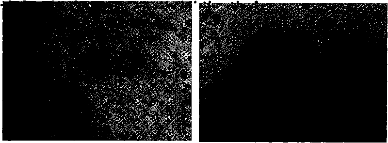
[0044] 2) Mix embryonic stem cells with endometrial cells in the form of single cells, add them to the collagen gel, adjust the pH to neutral with 0.1mol / L NaOH, and then add 10% Matrigel at a ratio of 4:1 to construct the embryo Stem cells are combined with the uterine sheet, and the differentiation and development of embryonic stem cells in the uterine sheet is observed (such as Figure 4 shown).

Embodiment 3

[0045] Example 3. Research on induced differentiation and development of embryonic stem cells:

[0046] In the established uterine differentiation and development model of embryonic stem cells, different inducers were added to observe the ability of embryonic stem cells to differentiate and develop into different adult cells. Figure 5 Shown is the detection of embryonic stem cells after they have formed terminally differentiated cells in a uterine sheet.


PUM

PropertyMeasurementUnit
diameteraaaaaaaaaa

Abstract

The invention relates to an artificial uterus three-dimensional gel model used for researching development and differentiation of embryonic stem cells, belonging to the research field of tissue engineering and regenerative medicine, and mainly solving the problem that no ideal bionic model exists for the research of differentiation and development of embryonic stem cells in vitro. The in vitro reconstructed tissue engineered uterus is adopted as a research model of the differentiation and development of the embryonic stem cells, wherein the embryonic stem cells are inoculated on the surface or evenly combined inside the uterus lamella, the development environment of normal embryos in vivo is simulated to regulate the microenvironment of the growth and development of the embryonic stem cells, and the reconstructed uterus tissues are taken as the main body to carry out the research on differentiation and development of embryonic stem cells, thus disclosing the inherent law and mechanism of the differentiation and development of embryonic stem cells in vitro, and searching for new methods for directional differentiation of embryonic stem cells to the functional cells of important vital organs at the terminal differentiation period; and the research results have an important meaning to disclose the development of the embryos in the early period, and have important theoretical meaning and application values for the curing based on embryonic stem cells.

Description

technical field [0001] The invention belongs to the research field of tissue engineering and regenerative medicine, and relates to a series of key technologies such as preparation of tissue-engineered uterus in vitro, embryonic stem cell culture and embryonic stem cell differentiation and development in vitro. This method can construct a tissue-engineered artificial uterine sheet or double-layered endometrium in vitro, and inoculate embryonic stem cells on the surface or evenly compound it inside to make it a research model for cell differentiation and development. By regulating the microenvironment of the growth of embryonic stem cells, the research on the differentiation and development of embryonic stem cells is carried out with the reconstructed uterine tissue as the main body, and then reveals the internal rules and mechanisms of embryonic stem cell differentiation and development in vitro, and seeks for embryonic stem cells to be in the terminal differentiation of importa...

Claims


Application Information

Patent Timeline
Patent Type & Authority Applications(China)
IPC IPC(8): A61L27/38A61L27/24
Inventor 王常勇周瑾郝彤段翠密林秋霞
Owner INST OF BASIC MEDICAL SCI ACAD OF MILITARY MEDICAL SCI OF PLA
Learn More