
Detection kit for distinguishing cow pathogenic mycobacteria infection from non-pathogenic mycobacteria infection and method thereof

The technology of Mycobacterium bovis and reagents is applied in the field of immunodetection, which can solve the problems of false positives, complex antigen components, and low sensitivity of serological detection methods, and achieve the effect of strong specificity and sensitivity

Active Publication Date: 2009-09-16
INST OF ANIMAL SCI OF CHINESE ACAD OF AGRI SCI
View PDF0 Cites 8 Cited by
  • Summary
  • Abstract
  • Description
  • Claims
  • Application Information

AI Technical Summary

Problems solved by technology

However, due to the complex antigenic components contained in PPD, some of the antigens are found in bovine pathogenic mycobacteria such as Mycobacterium tuberculosis and Mycobacterium bovis, and bovine non-pathogenic mycobacteria such as Mycobacterium avium and non-pathogenic environmental mycobacteria. Bacilli are widely present in the bacteria, resulting in poor detection specificity. At the same time, studies have shown that PPD cannot effectively distinguish between Mycobacterium bovis infection and BCG immunity, and false positives often occur in actual use.
[0006] Therefore, there is an urgent need to develop a detection reagent and method that can improve the specificity and sensitivity of detection at the same time, which can not only overcome the low sensitivity of serological detection methods, but also have a higher detection rate than PPD-stimulated IFN-γ release test. specificity

Method used


Image

  • Detection kit for distinguishing cow pathogenic mycobacteria infection from non-pathogenic mycobacteria infection and method thereof
  • Detection kit for distinguishing cow pathogenic mycobacteria infection from non-pathogenic mycobacteria infection and method thereof
  • Detection kit for distinguishing cow pathogenic mycobacteria infection from non-pathogenic mycobacteria infection and method thereof

Examples


Embodiment 1

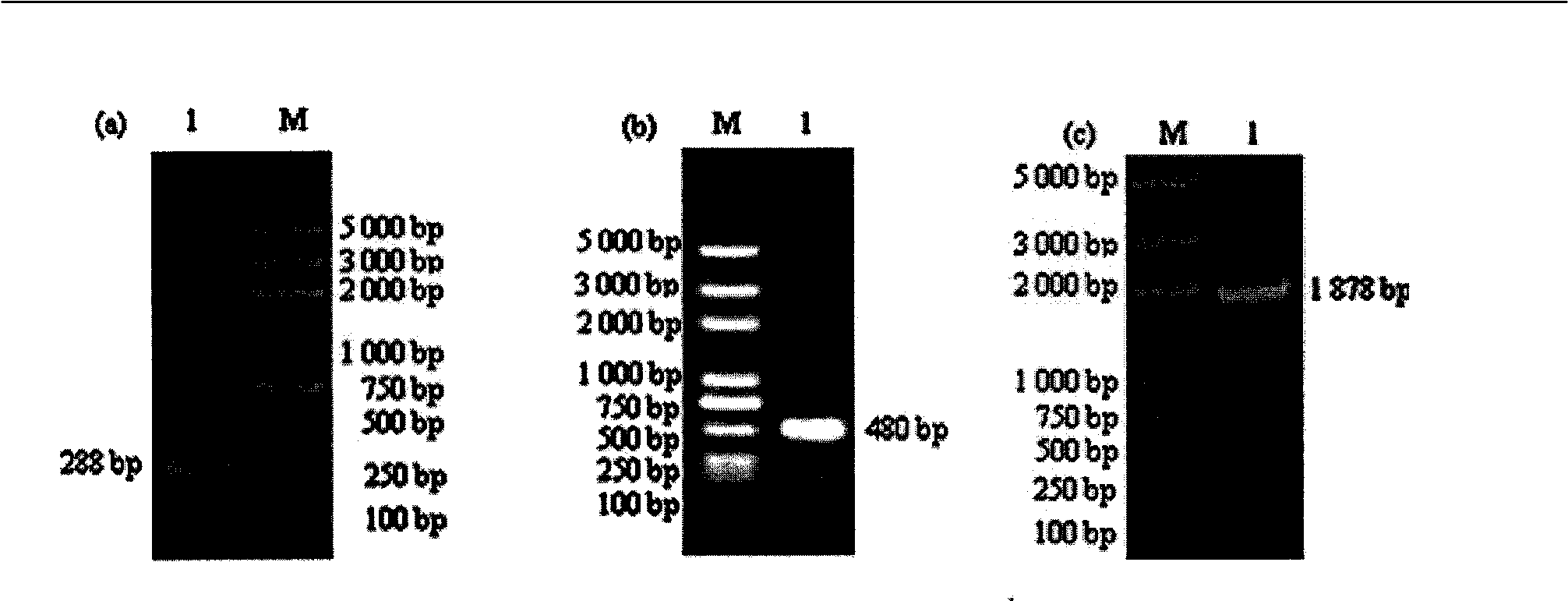
[0027] Example 1 Construction of recombinant plasmid pET-E6-M63-H70

[0028] 1.1 Extraction of Mycobacterium bovis genomic DNA

[0029] Refer to the method described in the instructions of the bacterial genomic DNA small amount rapid extraction kit (purchased from Beijing Broadtech Gene Technology Co., Ltd.).

[0030] 1.2 Design of primers

[0031] Specific primers were designed according to the ESAT-6, MPB63 and HSP70 gene sequences of Mycobacterium bovis genomic DNA (accession number BX248333) in GenBank. The primers were synthesized by Shanghai Shenggong Biotechnology Co., Ltd. The sequence is shown in Table 1 (the underlined is the enzyme Cut site, the shaded place is Linker).

[0032] Table 1 PCR primer name, sequence and size of amplified product

[0033]

[0034] 1.3 PCR amplification and product recovery of the target gene

[0035] Using M. bovis genomic DNA as a template, LA Taq DNA polymerase was used to amplify the ESAT-6, MPB63 and HSP70 genes. The specific reaction ...

Embodiment 2

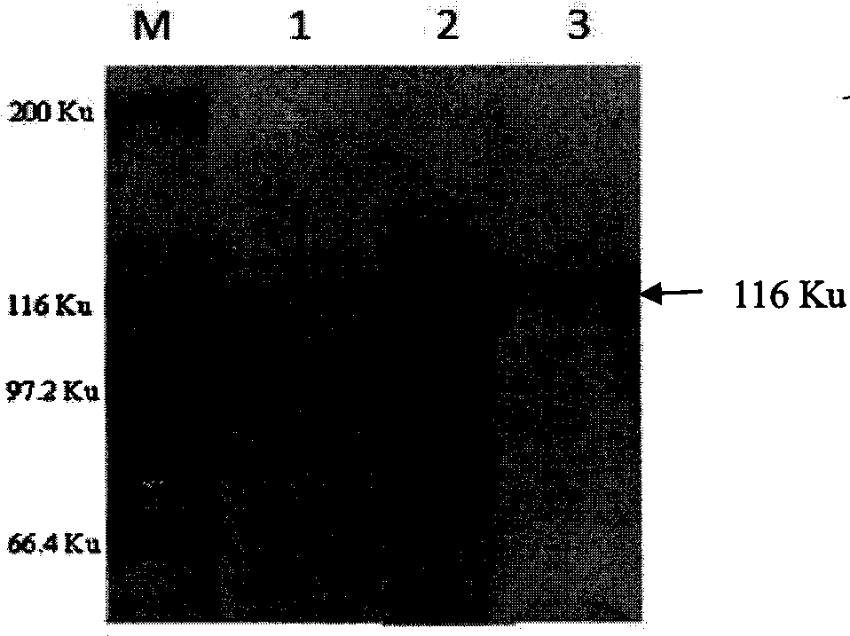
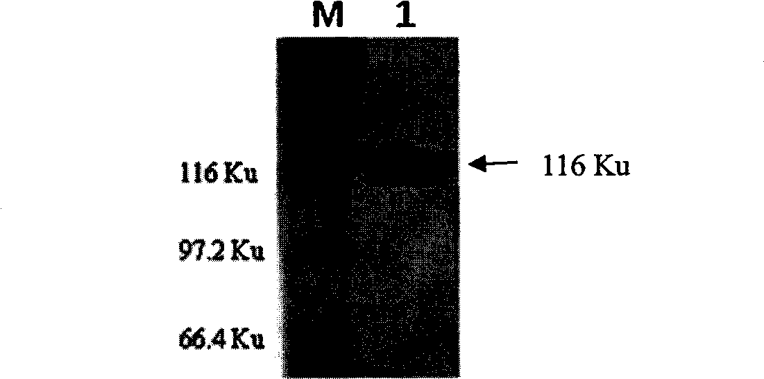
[0048] Example 2 Expression and purification of Mycobacterium bovis recombinant fusion protein rE6-M63-H70

[0049] 2.1 Induced expression, purification, SDS-PAGE and Western-blot analysis of recombinant fusion protein

[0050] The recombinant plasmid pET-E6-M63-H70 prepared in Example 1 was transformed into BL21(DE3) competent cells, and a single colony was picked and inoculated into 200 mL of LB medium containing a final concentration of 25 μg / ml ampicillin at 37°C. Incubate in bed to OD600 = 0.7, add IPTG with a final concentration of 1 mM, induce culture at 30°C, 160 rpm shaker for 10 hours, centrifuge at 9000 rpm for 10 minutes to collect the bacteria, resuspend the bacteria in 60 mL PBS (pH 7.4), and ultrasonically break the bacteria in an ice bath After crushing, the mixture was centrifuged at 12000 rpm, 4°C for 30 min, and then the supernatant was taken. Press His Link TM Protein Purification Resin (purchased from Beijing Zeping Technology Co., Ltd.) operation manual to pu...

Embodiment 3

[0052] Example 3 Establishment of detection method for Mycobacterium bovis

[0053] 3.1 Collection and cultivation of whole blood from cattle to be inspected

[0054] ① Collect 5ml of bovine heparin anticoagulant whole blood under aseptic conditions, transport it to the laboratory at room temperature (22±5℃) and culture within 8 hours after blood collection. ②Add anticoagulant blood to a 24-well tissue culture plate, 1.5ml / well, 2 wells for each cow to be inspected, and then sterilely add 50μl Tris-Cl (100mM, pH8.0, as a negative control stimulus), 50μl Put the solution (20μg) containing rE6-M63-H70 into the corresponding well, mix well, 37℃ CO 2 Incubate for 24h in an incubator. ③After incubation, carefully draw 400μl of upper plasma and transfer it into a 1.5ml centrifuge tube (plasma can be stored at 2-8°C for 7 days, -20°C for several months), and 50μl of plasma can be drawn from each tube as the sample to be tested. ELISA detection of the release level of bovine IFN-γ.

[005...


PUM


Abstract

The invention belongs to the field of immunodetection and relates to a detection kit for distinguishing cow pathogenic mycobacteria infection from non-pathogenic mycobacteria infection and a method thereof. The detection reagent comprises combined fusion protein rE6-M63-H70 used as a specific stimulation origin, the combined fusion protein can effectively stimulate sensitized peripheral blood lymphocyte cultured in vitro to release Gamma-interferon (IFN-Gamma) at a high level. The cow IFN-Gamma release test established by using the detection reagent rE6-M63-H70 combined fusion protein as the stimulation origin overcomes the insufficiencies of serology detection method and the IFN-Gamma release test with PPD as the stimulation origin, thus enjoying very high sensitivity and specificity and distinguishing cow pathogenic mycobacteria ( such as mycobacterium bovis) infection from non-pathogenic mycobacteria (such as mycobacterium avium or non-pathogenic mycobacteria) infection and even distinguishing the cow pathogenic mycobacteria infection from BGG immunity; therefore, the detection kit and the method of the invention can be effectively used to detect the clinical cow pathogenic mycobacteria infection.

Description

Technical field [0001] The invention belongs to the technical field of immune detection. The invention relates to a kit and a method for distinguishing between pathogenic and non-pathogenic mycobacterial infections in cattle. Background technique [0002] Bovine tuberculosis is a zoonotic chronic wasting infectious disease caused by Mycobacterium bovis and Mycobacterium tuberculosis. Cross infection between humans and animals has caused the disease to be widespread, so it has a very important public Sanitary significance. The World Health Organization (WHO) pointed out: "In countries where bovine tuberculosis exists, humans are always threatened. Unless bovine tuberculosis is eliminated, the control of human tuberculosis will not succeed." At present, some more developed countries and regions, such as the United States, Australia and Northern Europe have basically eliminated bovine tuberculosis. However, bovine tuberculosis is still one of the most common multiple diseases in my ...

Claims


Application Information

Patent Timeline
IPC IPC(8): G01N33/569G01N33/543C07K19/00
Inventor 朱鸿飞侯绍华郝辉贾红张泉毛开荣
Owner INST OF ANIMAL SCI OF CHINESE ACAD OF AGRI SCI
Learn More