Dental X-ray three-dimensional filming system and three-dimensional imaging method

A 3D image and dental technology, applied in the fields of dental radiology diagnosis, radiodiagnosis clinical application, etc., can solve the problem of not being able to provide 3D structure images of teeth, and achieve the effect of clear and delicate images, high safety, and high accuracy

Inactive Publication Date: 2009-04-29
王乔生
View PDF0 Cites 13 Cited by
  • Summary
  • Abstract
  • Description
  • Claims
  • Application Information

AI Technical Summary

Problems solved by technology

The real problem is that conventional dental X-ray cameras and dental X-ray panoramic cameras can only provide two-dimensional tooth structure images, but cannot provide three-dimensional structure images of teeth

Method used


Image

  • Dental X-ray three-dimensional filming system and three-dimensional imaging method

Examples


Embodiment Construction

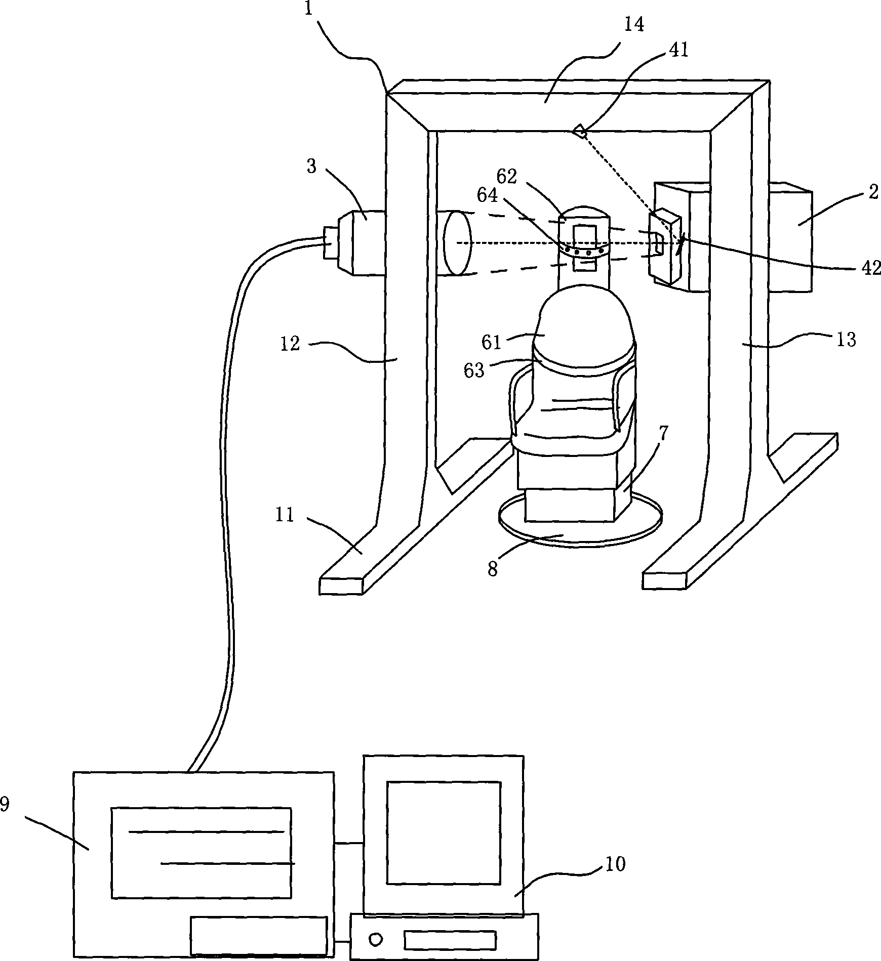
[0013] Such as figure 1 As shown, the dental X-ray three-dimensional imaging system provided in this embodiment includes: host gantry frame 1, X-ray generator head 2, X-ray detector 3, laser positioning device, additional mark 5, seat 6, seat Lifter 7, seat rotating device 8, image processing computer 9, operation and display interface 10. The host gantry frame 1 is composed of a base 11, two support arms 12, 13 and a beam 14. The X-ray generator head 2 and the X-ray detector 3 are relatively arranged on the two support arms 12, 13 of the host gantry frame. The requirement of the position of the laser positioning device is that the laser light produced by it coincides with the center line of the X-ray beam. In the present embodiment, the laser positioning device includes a laser source 41 and a reflection mechanism 42. The laser light emitted by the laser source 41 is kept in line with The centerlines of the X-ray bundles coincide. Seat 6 is arranged between two support arms...


PUM

PropertyMeasurementUnit
Rotation angleaaaaaaaaaa

Abstract

The invention discloses a dental X-ray three-dimensional imaging system and a corresponding three-dimensional imaging method, and aims at reconstructing a three-dimensional dental model image from a two-dimensional panorama dental X-ray image. The system comprises a host gantry frame, an X-ray generator head, an X-ray detector, a laser positioning device, an attaching sign, a chair, a chair riser, a chair rotating device, an image processing computer and an operation and display panel. The method comprises: (1) teeth are carried out with panoramic X-ray imaging by rotating the chair; (2) pre-imaging process is carried out with the image processing computer; and (3) the three-dimensional image is reconstructed with the image processing computer. The system and the method take a digital panoramic camera regularly used in dental operation as the main part to realize the optimal cone-typed beam three-dimensional image reconstruction by integrating the optical laser positioning device. The system is characterized by high safety and precision, convenient operation, clear and fine picture, good practicability and durability, etc.

Description

technical field [0001] The invention relates to an X-ray imaging system and an imaging method in the medical field. Background technique [0002] Computer-assisted surgical treatment has been widely used in medicine. In clinical dental surgery, there is also an urgent need for three-dimensional three-dimensional image positioning aids. Such as orthodontics, implants, teeth, periodontal, maxillofacial surgery, dental cosmetology, teaching and scientific research, etc. in dental surgery, all need the assistance of computer images to realize the planning and guidance of dental surgery, and use this Improve the quality of surgery while reducing the risk of surgical treatment. [0003] In actual dentistry, it is common to use two-dimensional X-ray dental photographs to guide the planar structure of dental surgery. In the complex dental implant process, doctors often need to use composite 2D / 3D images of teeth, and more importantly, rely on very important Dental bone density dat...

Claims


Application Information

Patent Timeline
IPC IPC(8): A61B6/14
Inventor 王乔生
Owner 王乔生
Why Eureka
Try Eureka