Eureka AIR delivers breakthrough ideas for toughest innovation challenges, trusted by R&D personnel around the world.

Three-dimensional stephanoporate organization engineering bracket material, fibre cementing method preparing same and applications thereof

A tissue engineering scaffold, a three-dimensional porous technology, is used in medical science, prostheses, bone implants, etc. to achieve the effects of stable scaffold system structure, simple preparation process and uniform pore structure.

Inactive Publication Date: 2008-08-27
DONGHUA UNIV
View PDF3 Cites 18 Cited by
  • Summary
  • Abstract
  • Description
  • Claims
  • Application Information

AI Technical Summary

Problems solved by technology

[0008] The purpose of the present invention is to provide a three-dimensional porous tissue engineering scaffold material, its preparation and application by fiber bonding method, the present invention combines the advantages of existing biological materials, overcomes the deficiency in the performance of a single material, and prepares a tissue engineering scaffold material With good biocompatibility, sufficient mechanical properties, and adjustable degradation rate, it can be used as a bone or cartilage tissue defect repair material and a cell scaffold material for in vitro tissue culture, which can meet the needs of the development of a new generation of biomaterials

Method used


Image

  • Three-dimensional stephanoporate organization engineering bracket material, fibre cementing method preparing same and applications thereof

Examples


Embodiment 1

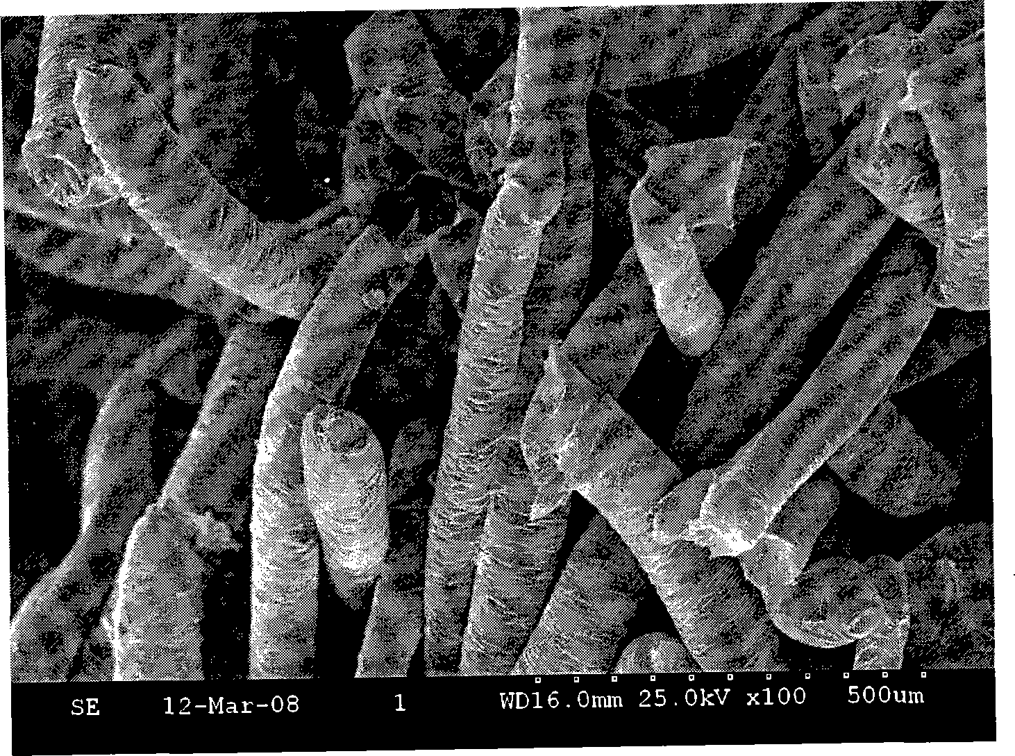
[0025] The high polymer PBS with a molecular weight of 40,000 and a melting point of 115°C, and the high polymer PCL with a molecular weight of 80,000 and a melting point of 63°C were melt-spun at 130°C to obtain fibers with an average diameter of 300 μm, which were cut into After the equal length is 8.0mm, fill it into a mold with a specific shape. The filling quality is calculated based on the porosity of 85%. After demoulding, the tissue engineering carrier material with specific shape and good performance can be obtained. The material has a pore size of 160-400 μm measured by a scanning electron microscope, a porosity of 80% measured by a liquid displacement method, and a degree of pore penetration of more than 90%. The in vitro degradation cycle is appropriate and the mechanical properties are excellent. The material can be used as a bone tissue engineering carrier material for repairing and reconstructing bone damage.

Embodiment 2

[0027] The high polymer PBS with a molecular weight of 80,000 and a melting point of 115°C, and the high polymer PCL with a molecular weight of 100,000 and a melting point of 63°C were melt-spun at 140°C to obtain fibers with an average diameter of 100 μm, which were cut into After the equal length is 10.0mm, fill it into a mold with a specific shape. The filling quality is calculated based on the porosity of 85%. After demoulding, the tissue engineering carrier material with specific shape and good performance can be obtained. The material has a pore diameter of 70-200 μm measured by a scanning electron microscope, a porosity of 85% measured by a liquid displacement method, and a degree of pore penetration of more than 90%. The in vitro degradation cycle is appropriate and the mechanical properties are excellent. The material can be used as a bone tissue engineering carrier material for repairing and reconstructing bone damage.

Embodiment 3

[0029] The high polymer PBS with a molecular weight of 100,000 and a melting point of 118°C, and the high polymer PCL with a molecular weight of 200,000 and a melting point of 64°C were melt-spun at 150°C to obtain fibers with an average diameter of 140 μm, which were cut into After the equal length is 8.0mm, fill it into a mold with a specific shape. The filling quality is calculated based on the porosity of 85%. The temperature of the vacuum oven is set at 80°C. After demoulding, the tissue engineering carrier material with specific shape and good performance can be obtained. The material has a pore size of 100-200 μm measured by a scanning electron microscope, a porosity of 81% measured by a liquid displacement method, and a degree of pore penetration of more than 90%. The in vitro degradation cycle is appropriate and the mechanical properties are excellent. The material can be used as a bone tissue engineering carrier material for repairing and reconstructing bone damage. ...


PUM

PropertyMeasurementUnit
melting pointaaaaaaaaaa
pore sizeaaaaaaaaaa

Abstract

The invention relates to a three-dimensional porous tissue engineering stent material, a fiber-gluing preparation method thereof and the application thereof, The material comprises poly butylene succinate and polycaprolactone; the weight portion is: poly butylene succinate 90 to 10 parts, polycaprolactone 10 to 90 parts; preparation: 1) mixing poly butylene succinate and polycaprolactone to obtain fiber by melt spinning; 2) cutting fiber and filling the fiber into a module, the fiber being arranged in an even structure; 3) putting the mould into a vacuum oven with 50 to 90 DEG C and keeping for 5 minutes to 1 hour, demoulding at room temperature and drying in vacuum after cooling, and obtaining three-dimensional tissue engineering stent material; 4) sterilizing and packing the three-dimensional porous tissue engineering stent material; The material has the application of being used as bone or cartilage tissue engineering cytoskeleton for repairing and rebuilding of bone or cartilage tissue organ. The tissue engineering stent material has evenly structured, internal pores that communicate with each other. The aperture of the pores is 10 to 500 Mum, and the porosity changes between 70 to 91 percent, so that the systematic structure of the stent is stable and easy to produce.

Description

technical field [0001] The invention belongs to the field of tissue engineering scaffold material and its preparation, in particular to a three-dimensional porous tissue engineering scaffold material, its fiber bonding method preparation and application. Background technique [0002] In daily life, the loss or failure of human tissues and organs is very common, which poses a serious threat to people's health and life. For a long time, human beings have been exploring and researching the use of materials and biotechnology to improve their own health. Traditional treatment methods include tissue and organ transplantation, surgical reconstruction, drug therapy, treatment with artificial substitutes and mechanical devices, etc., but these methods all have their obvious shortcomings. In the 1990s, with the development of cell biology, molecular biology, material science and related physical and chemical disciplines, tissue engineering was proposed as a new treatment method, and ...

Claims


Application Information

Patent Timeline
Patent Type & Authority Applications(China)
IPC IPC(8): A61L27/18A61L27/56A61F2/28
Inventor 杨庆陈思诗郯志清沈新元彭兰兰
Owner DONGHUA UNIV
Why Eureka
Eureka Blog
Learn More