Method and apparatus for increasing field of view in cone-beam computerized tomography acquisition

a computerized tomography and field of view technology, applied in tomography, instruments, applications, etc., can solve the problems of insufficient compensation and more expensive components, and achieve the effect of reducing patient movement, shortening acquisition time, and reducing the cost of components

Active Publication Date: 2017-10-24
CEFLA SOC COOP
View PDF6 Cites 145 Cited by
  • Summary
  • Abstract
  • Description
  • Claims
  • Application Information

AI Technical Summary

Benefits of technology

[0025]A first advantage of the present invention consists in providing an apparatus that, the cost of components being equal, can provide reconstructed volumes bigger than what is obtainable with the same X-ray detector without EV protocol.
[0026]A second advantage consists in the shortening of acquisition time, through the performance of two or more consecutive subacquisitions, minimizing patient's movements. The repositioning of ray group with respect to the patient occurs automatically, keeping the patient still. In this way both motion artifacts and registration volumes are minimized.
[0027]A third advantage lies in the fact that with the present invention radio-opaque markers are not necessary, in that registration is performed on the basis of the anatomic elements present in the volumes to be registered.
[0028]A fourth advantage consists in that the volumes are directly registered, without need of volume pre-processing.
[0029]A fifth advantage consists in the parallelization of the registration algorithm, which allows reducing the computation times.

Problems solved by technology

Generally speaking, each of these solutions involves significant disadvantages: e.g. economic disadvantages, in that the components are more expensive, or otherwise ergonomic disadvantages, linked to the need to increase the overall dimensions of the apparatus.
As illustrated, the effect of the compensation does not fully recover the lack of homogeneity of the distribution of the radiation along the active area of the sensor.
Another drawback can be seen in comparing the scanning process of a traditional CBCT configuration with the one using an offset sensor (FIGS. 2 and 3a to 3c of U.S. Pat. No. 8,363,780).
Thus in cases where the extended FOV obtained by the sensor's offset is still insufficient for covering the entire region of interest, U.S. Pat. No. 8,363,780 does not suggest a solution for further expanding the FOV so that it covers the region of interest.

Method used


Image

  • Method and apparatus for increasing field of view in cone-beam computerized tomography acquisition
  • Method and apparatus for increasing field of view in cone-beam computerized tomography acquisition
  • Method and apparatus for increasing field of view in cone-beam computerized tomography acquisition

Examples


first embodiment

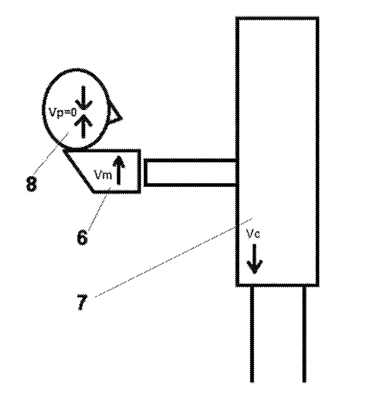
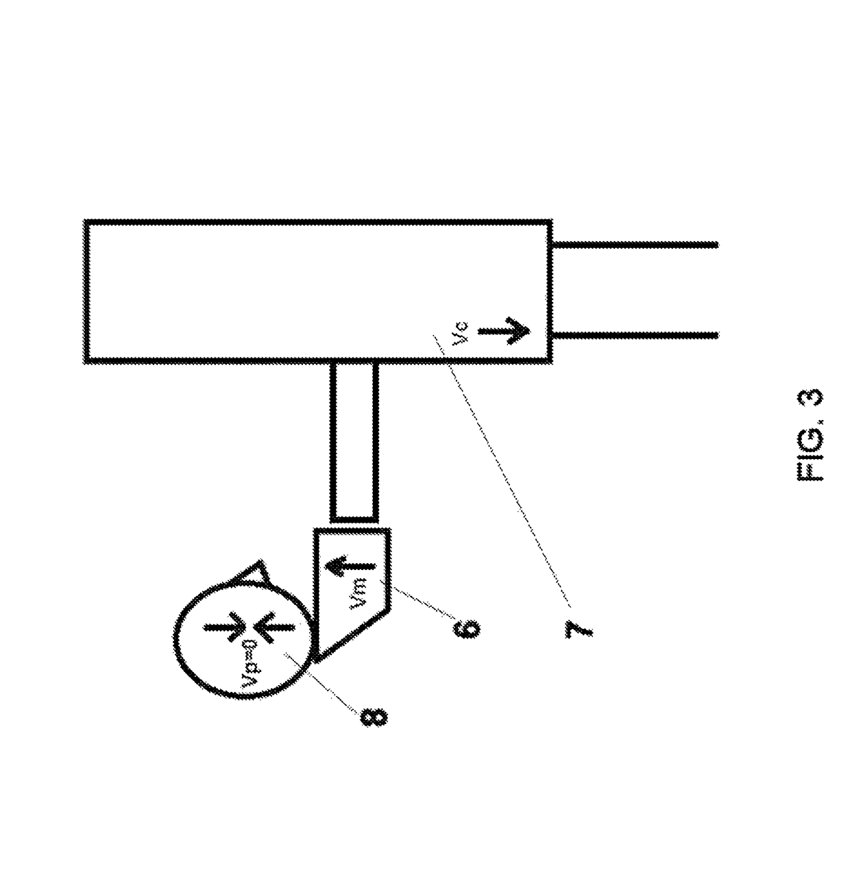
[0042]In a first embodiment, the present invention is integrated in the vertical apparatus shown in FIG. 2.

[0043]In this first embodiment, patient 8 is positioned in patient positioning group 6, and a first subacquisition of a first subvolume is performed (e.g. of the patient's mandible). At the end of this first subacquisition, either ray group 4 or patient positioning group 6 is re-positioned and a second subacquisition is performed (e.g. of the patient's mandible). Once the two subvolumes have been acquired, they are registered so as to obtain a reconstructed FOV bigger than that obtainable through a single acquisition, as described in the flow chart of FIG. 8.

[0044]According to the desired result, a vertical addition (e.g. mandible+maxilla) or a horizontal addition (e.g. right half of arch+left half of arch) may be necessary.

[0045]If a vertical volume addition is desired, the repositioning between the subacquisitions occurs with a synchronized movement of the lifting post and of...

second embodiment

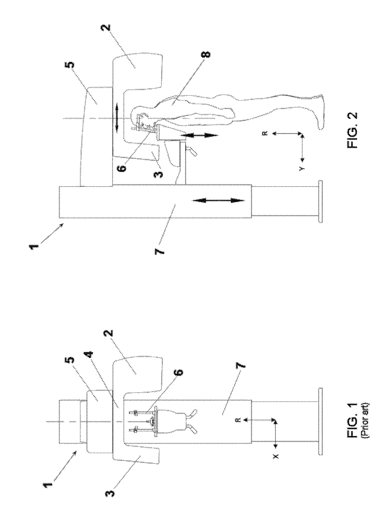
[0052]A second embodiment relates to the horizontal apparatus shown in FIG. 5.

[0053]If a vertical volume addition must be performed, (longitudinal with respect to patient's long axis), the patient is suitably positioned on table 16, and a first subacquisition of a first subvolume is performed (e.g. of the patient's mandible). At the end of this first subacquisition, table 16 is repositioned and a second subacquisition is performed to acquire a second subvolume (e.g. of the patient's maxilla). Once the two subvolumes have been acquired, they are registered so as to obtain a reconstructed FOV bigger than that obtainable through a single acquisition, as described in the flow chart of FIG. 8. In particular, in this case table 16 is translated at a suitable distance along axis 21, perpendicular to the plane of gantry 14. An example of the result of such vertical volume addition is shown in FIG. 6.

[0054]If a horizontal volume addition is required (perpendicular to patient's long axis), th...


PUM


Abstract

A method and apparatus for Cone-Beam Computerized Tomography, (CBCT) is configured to increase the maximum Field-Of-View (FOV) through a composite scanning protocol and includes acquisition and reconstruction of multiple volumes related to partially overlapping different anatomic areas, and the subsequent stitching of those volumes, thereby obtaining, as a final result, a single final volume having dimensions larger than those otherwise provided by the geometry of the acquisition system.

Description

FIELD OF THE INVENTION[0001]The present invention relates to a method and an apparatus for Cone-Beam Computerized Tomography (CBCT) that increases the maximum Field-Of-View (FOV) through a composite scanning protocol, based on the acquisition and reconstruction of multiple volumes by partially overlapping different anatomic areas, and subsequently stitching said volumes, thereby obtaining, as a final result, a single final volume having dimensions larger than those otherwise allowed by the geometry of the acquisition system.BACKGROUND OF THE INVENTION[0002]CBCT apparatuses are known in the art and provide tomographic images of an anatomic portion by acquiring a sequence of bi-dimensional radiographic images during the rotation of a system that comprises an X-ray source and an X-ray detector around the anatomic part to be imaged.[0003]A CBCT apparatus includes substantially: an X-ray source projecting a conic X-ray beam (unless it is subsequently collimated) through an object to be a...

Claims


Application Information

Patent Timeline
Patent Type & Authority Patents(United States)
IPC IPC(8): A61B6/00G06T3/40A61B6/03G06T11/00A61B6/14
CPCA61B6/5264A61B6/032A61B6/4085A61B6/5211A61B6/5241G06T11/008G06T3/4038G06T2200/32A61B6/14A61B6/5205A61B6/54G06T2200/04
Inventor MENEGAZ, GLORIAMORSIANI, ROBERTORIVALTA, BRUNOSANDRI, CARLOSIMONETTI, FLAVIO
Owner CEFLA SOC COOP
Why Eureka
Try Eureka