Implants and biodegradable tissue markers
a biodegradable and tissue-based technology, applied in the field of tissue gaps stabilization and visualization, can solve the problems of achieve the effects of preventing targeting, good visualization of margins, and poor resolution of site margins
- Summary
- Abstract
- Description
- Claims
- Application Information
AI Technical Summary
Benefits of technology
Problems solved by technology
Method used
Image
Examples
example 1
Conformal Filling with Hydrogels
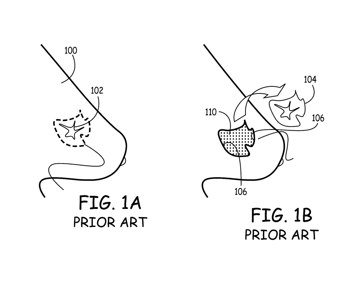
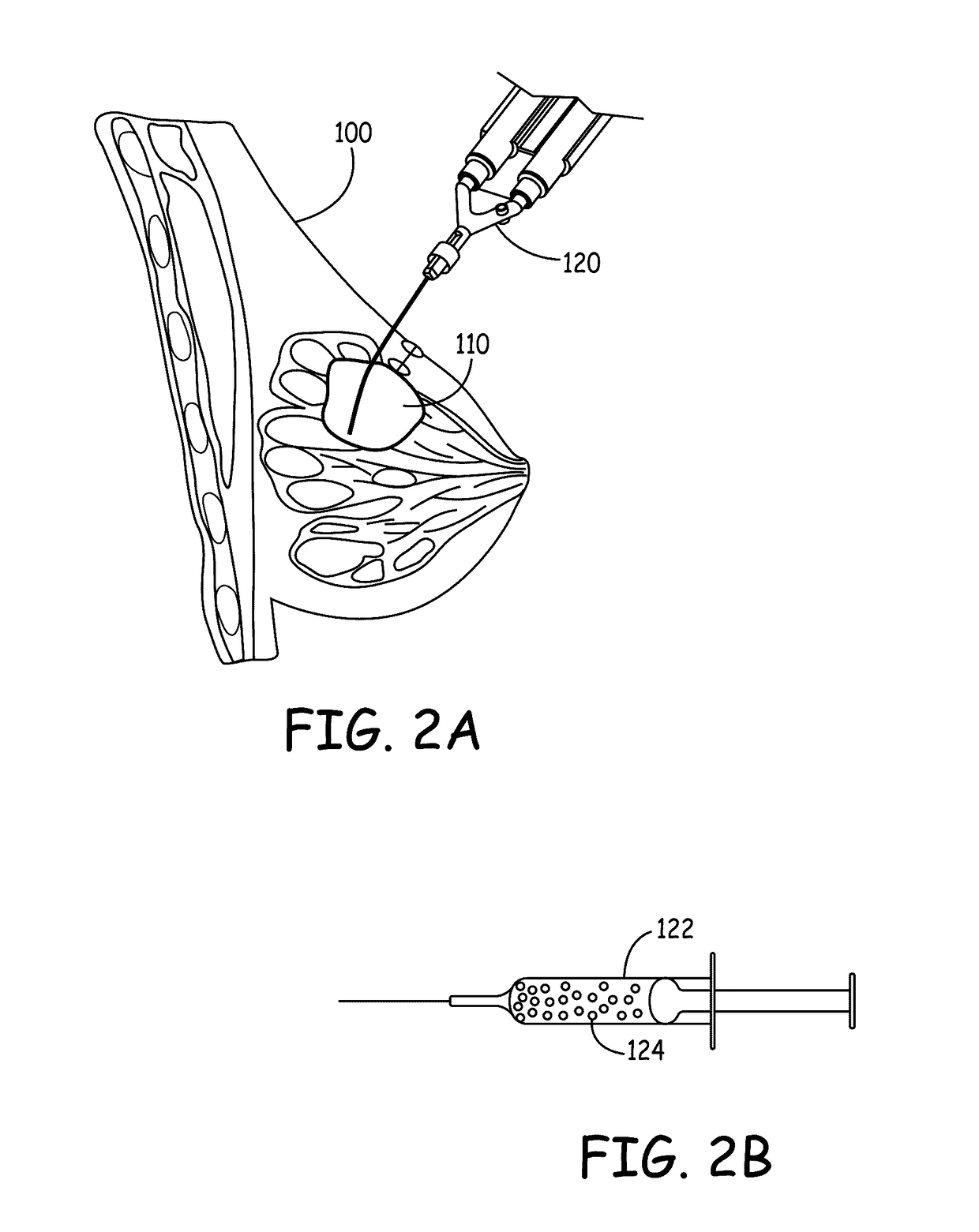
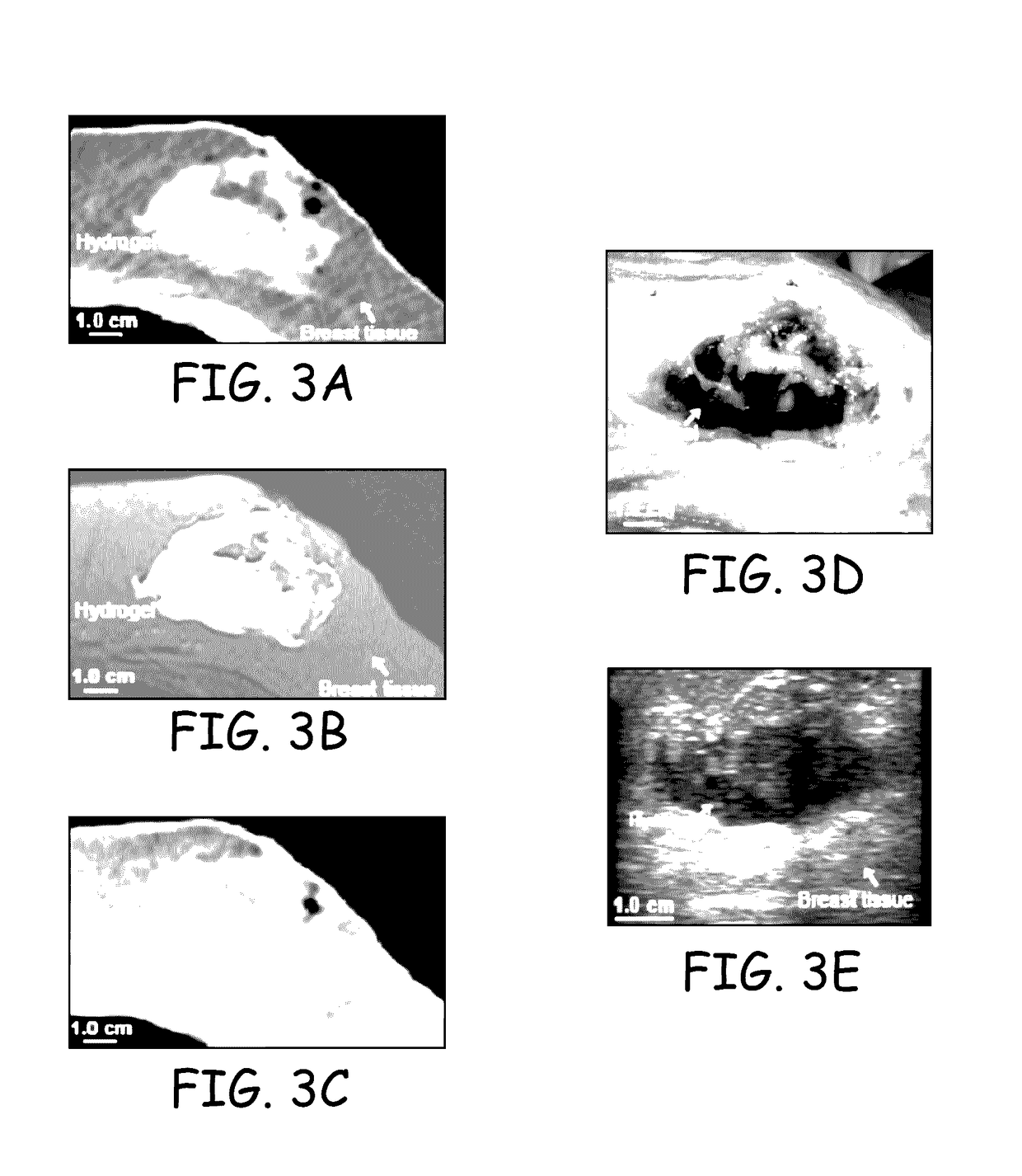
[0152]Under an approved protocol, three cadaver specimens were obtained for bilateral lumpectomies; in one, unilateral lumpectomy was performed due to prior breast surgery. On the CT-simulation table, each specimen was positioned for left sided lumpectomy (using wedges). Lumpectomies ranging from 31 to 70 cc were performed. Following lumpectomy, a 0.25″ diameter silicone catheter was placed within the cavity, the subcutaneous tissue was apposed, and the skin was closed. In one case, the superior and inferior cavity walls were apposed prior to closure. Before hydrogel injection, each underwent CT simulation (Philips BRILLIANCE BIG BORE CT, 3 mm slices, 120 kVp, 300 mA, 60 cm FOV).
[0153]Following CT simulation, 18 to 70 cc of hydrogel was injected within the cavity, the silicone catheter was withdrawn, and the hydrogel was allowed to solidify. The hydrogel, when injected, has the viscosity of water but then, within 60 seconds, polymerized and formed a s...
example 2
Radiation Exposure Control with Conformal Hydrogels
[0157]The specimens of Example 1 had radiation plans developed for pre-hydrogel implant cases and post-hydrogel implant cases. The pre-hydrogel and post-hydrogel CT scans were imported into a Pinnacle treatment planning system (v8.0m, Philips Radiation Oncology Systems). For first sets of plans, standard margin expansions (a 15 mm GTV-CTV expansion and a 10 mm CTV-PTV expansion) was used for all five pre-hydrogel and post-hydrogel plans (per the NSABP-B-39 / RTOG-0413 protocol). For a second set of plans, standard margins were used for the prehydrogel plans, but reduced margins (a 10 mm GTV-CTV expansion and a 5 mm CTV-PTV expansion) were used for the post-hydrogel plans.
[0158]Intensity modulated radiotherapy (IMRT) treatment planning was performed using five, non-coplanar beams designed to minimize normal structure radiation exposure. Non-target structures included the ipsilateral and contralateral lungs, the heart, and the ipsilater...
example 3
Radioopaque Hydrogels and Imaging
[0161]The radiopacity of serial diluted Iohexol (OMNIPAQUE) was measured to obtain a CT number as a function of iodine concentration. A CT number (also referred to as a Hounsfield unit or number) is the density assigned to a voxel in a CT (computed tomography) scan on an arbitrary scale on which air has a density −1000; water, 0; and compact bone +1000. The CT number of diluted Iohexol ranged from 2976 at a 50% concentration, to 37 at a 0.2% concentration. These corresponded to a plot of iodine concentration versus CT number, with an iodine concentration of approximately 0.15% resulting in a CT number of about 90 (FIG. 8). Two different matrix formulations containing iodine at different concentrations were tested. First, iodine with succinimidyl glutarate (SG or SGA) functional terminal groups were complexed to 5000 Dalton linear PEG to make a PEG molecule complexed with iodine (PEG-I). Second, potassium iodide (KI) was incorporated into the gel at d...
PUM
 Abstract
Description
Claims
Application Information
 - R&D
- Intellectual Property
- Life Sciences
- Materials
- Tech Scout
- Unparalleled Data Quality
- Higher Quality Content
- 60% Fewer Hallucinations
Browse by: Latest US Patents, China's latest patents, Technical Efficacy Thesaurus, Application Domain, Technology Topic, Popular Technical Reports.
© 2025 PatSnap. All rights reserved.Legal|Privacy policy|Modern Slavery Act Transparency Statement|Sitemap|About US| Contact US: help@patsnap.com


