
Bioassay method for detecting physiologically active substance

a biological test and substance technology, applied in the field of biological test methods for detecting physiologically active substances, can solve the problems of poor sensitivity of the competition test method, limitations, and inability to detect avp in plasma with high sensitivity, and achieve the effect of simple and safe procedures

Active Publication Date: 2016-04-19
OTSUKA PHARM CO LTD
View PDF15 Cites 2 Cited by
  • Summary
  • Abstract
  • Description
  • Claims
  • Application Information

AI Technical Summary

Benefits of technology

The present invention allows for the safe and easy measurement of biological substances that affect the body's natural response to a molecule called cAMP. This is done without the need for complicated procedures involving radioisotopes.

Problems solved by technology

However, the ELISA method using the competition technique has poor sensitivity and has limitations in the assay of a microscale sample in blood.
Since AVP is a low-molecular-weight peptide, AVP in plasma cannot be assayed with high sensitivity due to the unavailability of sandwich ELISA assay using sandwich antibodies against a plurality of antigen determinants that permit higher sensitivity.

Method used


Image

  • Bioassay method for detecting physiologically active substance
  • Bioassay method for detecting physiologically active substance
  • Bioassay method for detecting physiologically active substance

Examples


examples

A. Experiment on Cell Expressing V2R

1. Details on Method for Constructing Frozen Cell

[0210]The cDNA sequence of human V2R (Genbank No. NM_001146151.1) (SEQ ID NO: 5) was amplified by the PCR method from a human-derived cDNA library and cloned into pUC18. The human vasopressin receptor isoform 2 (V2R) cDNA cloned in pUC18 was recloned into a pEF2 vector to prepare pEFhV2R.

[0211]The cDNA sequence of a cyclic nucleotide dependent calcium channel (Genbank No. BC048775) (SEQ ID NO: 4) was amplified by the PCR method from a mouse olfactory epithelial cell-derived cDNA library (1994-bp) and cloned into an expression vector (pCMVSPORT, Invitrogen Corp.) to prepare pmCNGα2 (FIG. 18). Furthermore, for enhancing cAMP selectivity and sensitivity thereto, a construct pmCNGα2MW expressing a modified cyclic nucleotide dependent calcium channel (SEQ ID NO: 6) in which 460th cysteine (C) is substituted with tryptophan (W) and the 583rd glutamic acid (E) is substituted with methionine (M) was prepare...


PUM

PropertyMeasurementUnit
timeaaaaaaaaaa
concentrationaaaaaaaaaa

Abstract

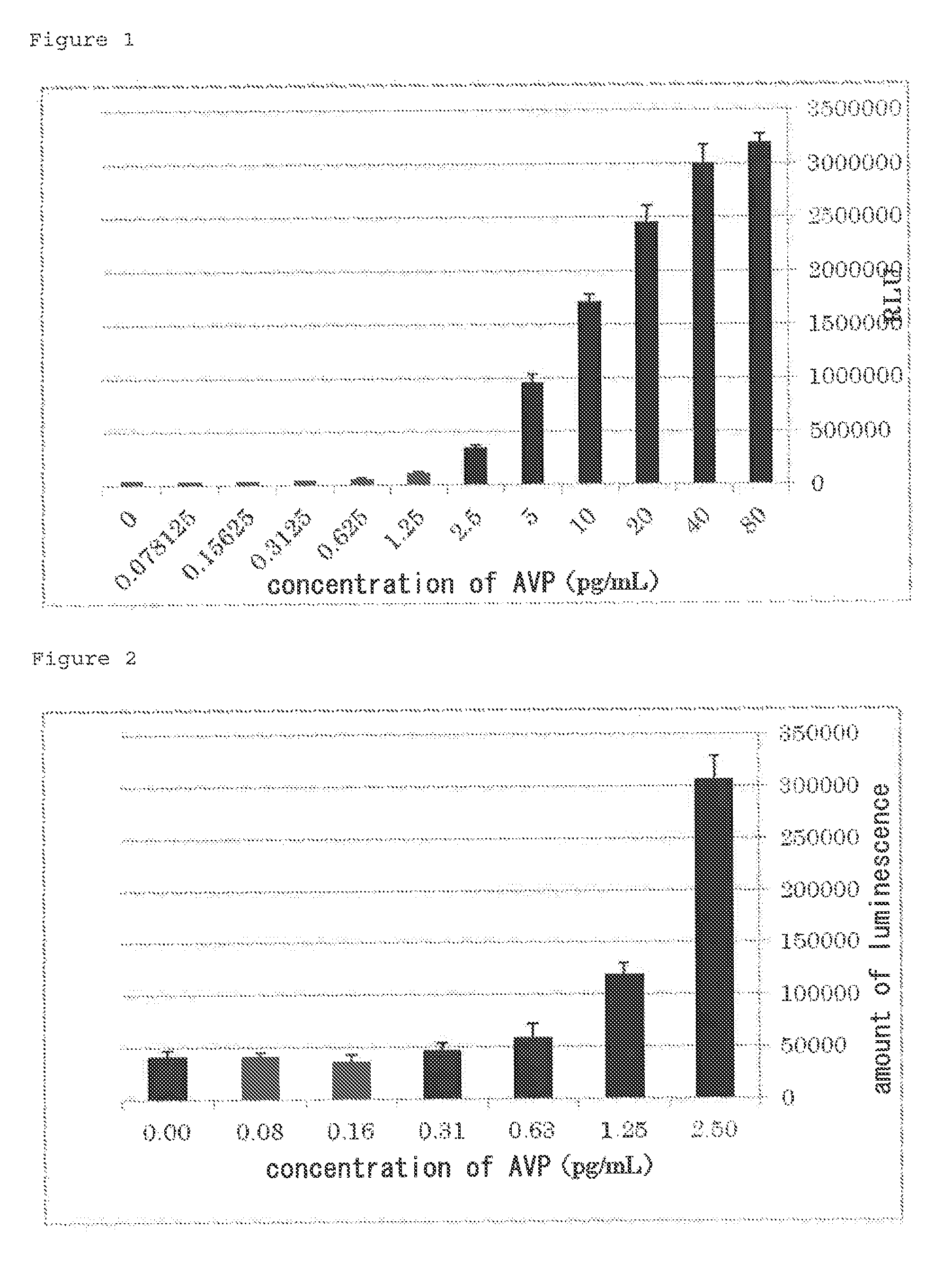
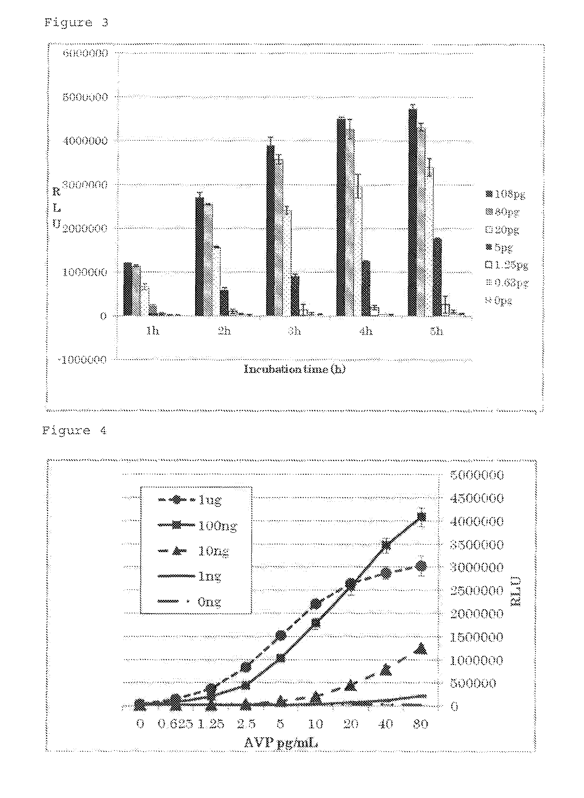
Provided are a procedurally simple and safe method and kit for measuring the concentration in a biological sample of a physiologically active substance which binds to a receptor that causes a change in intracellular cAMP concentration. By providing a composition including genetically-modified cells which show co-expression of a receptor that causes a change in intracellular cAMP concentration, a cyclic nucleotide-responsive calcium channel, and aequorin luminescent protein, it is possible to measure how much of a physiologically active substance which binds to said receptor is included in a biological sample.

Description

TECHNICAL FIELD[0001]The present invention relates to a cell expressing a receptor that alters intracellular cAMP concentration, a cAMP dependent calcium channel and a calcium sensitive protein, a composition comprising the cell, use of the composition for diagnosing a disease associated with a physiologically active substance that binds to the receptor, and a method for diagnosing the disease using the composition, etc.BACKGROUND ART[0002]Receptors to which physiologically active substances bind may serve as drug development targets. Particularly, receptors that alter intracellular cAMP concentration as second messengers, such as G protein-coupled receptors (hereinafter, also referred to as GPCRs), are important as drug development targets. Many agents that bind to such receptors have been developed so far. Examples of such GPCRs include receptors to which arginine vasopressin (hereinafter, also referred to as AVP) or glucagon-like peptide-1 (hereinafter, also referred to as GLP-1)...

Claims


Application Information

Patent Timeline
Patent Type & Authority Patents(United States)
IPC IPC(8): G01N21/64G01N33/68
CPCG01N21/6486G01N33/6872G01N2333/726G01N2500/10G01N2800/042C12N5/10C12N15/00C12N15/09
Inventor ARAKI, NAOHIROIIDA, MITSURUMACHIDA, KIYOTAKA
Owner OTSUKA PHARM CO LTD
Learn More