Dendritic cell modulation in post-ischemic wounds
a technology of dendritic cells and wounds, applied in the field of dendritic cell modulation in post-ischemic wounds, can solve the problems of increasing the risk of myocardial rupture following myocardial surgery, and achieve the effects of promoting post-ischemic wound healing, reducing mi size, and preserving left ventricular function
- Summary
- Abstract
- Description
- Claims
- Application Information
AI Technical Summary
Benefits of technology
Problems solved by technology
Method used
Image
Examples
example 1
Anti-DC Immunotoxins for Use in Mice and Humans
[0163]A PE38-based anti-VLC immunotoxin to target DC in mice was prepared using a truncated mutant of Pseudomonas aeruginosa exotoxin A (PE38) that has exhibited great safety in clinical trials. Kreitman et al. (2005) J. Clin. Oncol. 23:6719-29. The two variable chains (VH and VL) of the anti-CD11c hybridoma N418 (ATCC #HB-224). The sequences were then connected in frame by a fragment encoding for a peptide linker, as described. Demangel et al. (2005) Mol. Immunol. 42:979-985. Next, this anti-CD11c scFv was subcloned in frame with the hinge sequence of CD8, followed by PE38 (ATCC #67205). The exotoxin encoded by this sequence lacks the cell-binding domain, so the protein is harmless in the absence of a recognition sequence. However, the single-chain antibody in the resulting fusion gene binds to CD11c. It is therefore internalized by CD11c+ cells, bringing the lethal activity to the cytosol of VLCs. Kreitman et al. (2005) J. Clin. Oncol...
example 2
Appearance of Vascular Leukocytes Following Myocardial Infarction
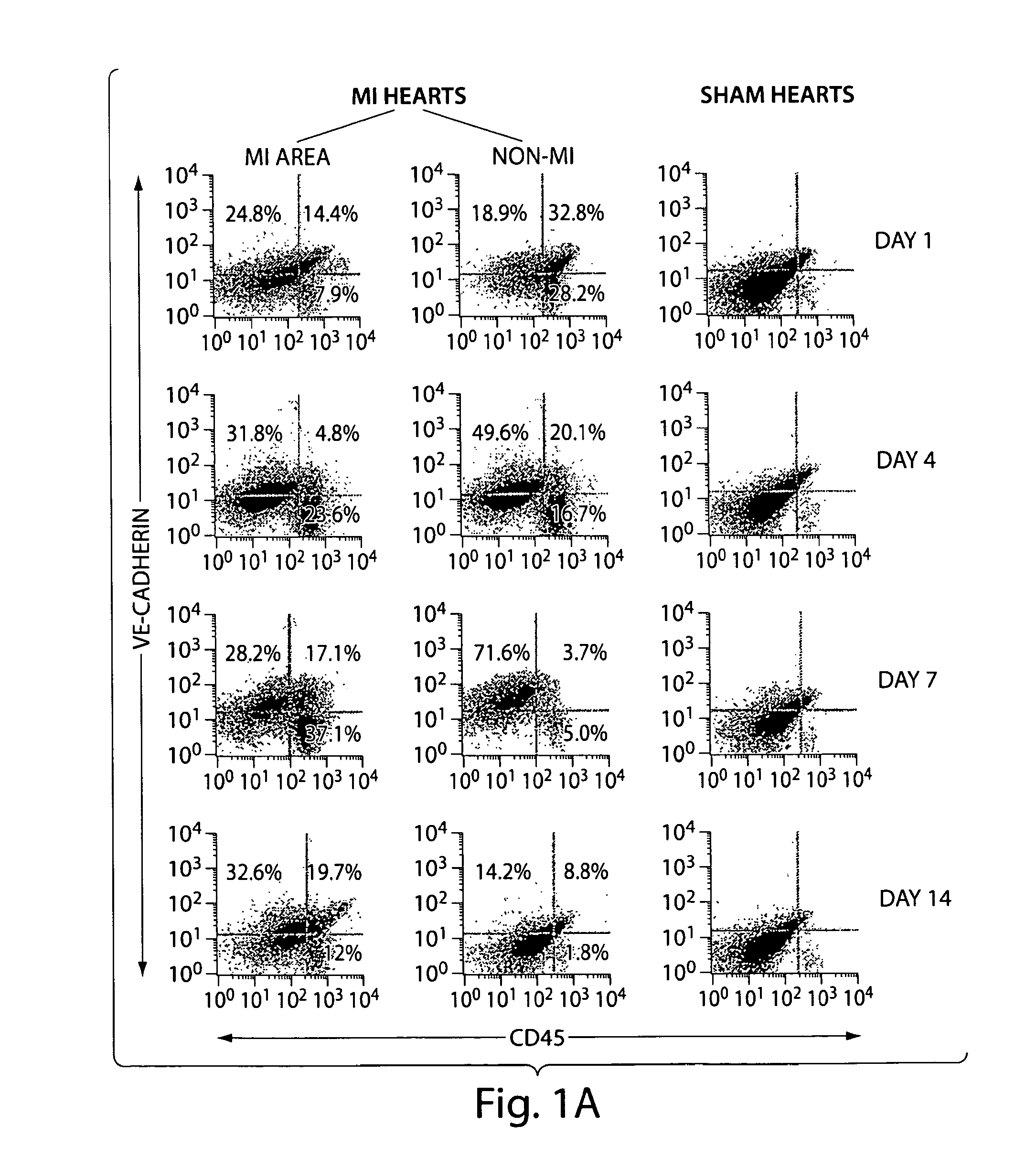
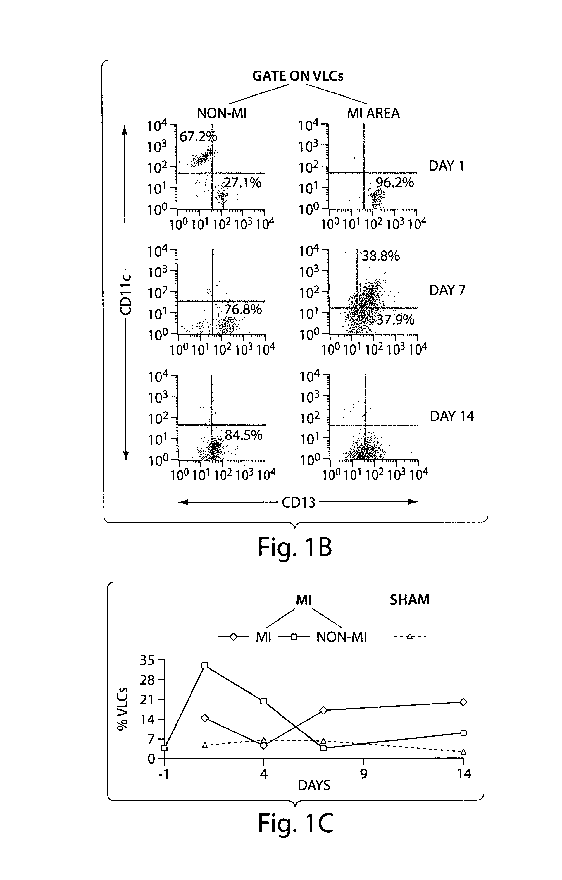
[0166]In a time course experiment of MI in wild type mice, vascular leukocytes (VLC), i.e. cells that express both leukocyte and endothelial markers, appeared in the viable segment of the hearts as early as two days after induction of MI. At four days after MI these cells are located in the vessels of the healing infarct wound, and they remain present in these vessels up to 14 days after MI. At this latter time point VLC were also found in the extravascular space of the infarct area.
[0167]The resolution of light microscopy and confocal microscopy was not sufficient to determine the exact location of VLC relative to the endothelial cells of the vasculature in the healing MI wound. Electron microscopy at four days post-MI revealed that dendritic cells are located at the abluminal surface of the basement membrane in vessels of the infarct wound, in close proximity to the endothelial cells.
[0168]FIG. 1 summarizes the resul...
example 3
Depletion of CD11c+ Dendritic Cells During Wound Healing after Myocardial Infarction Significantly Reduces Infarct Size and Preserves LV Function
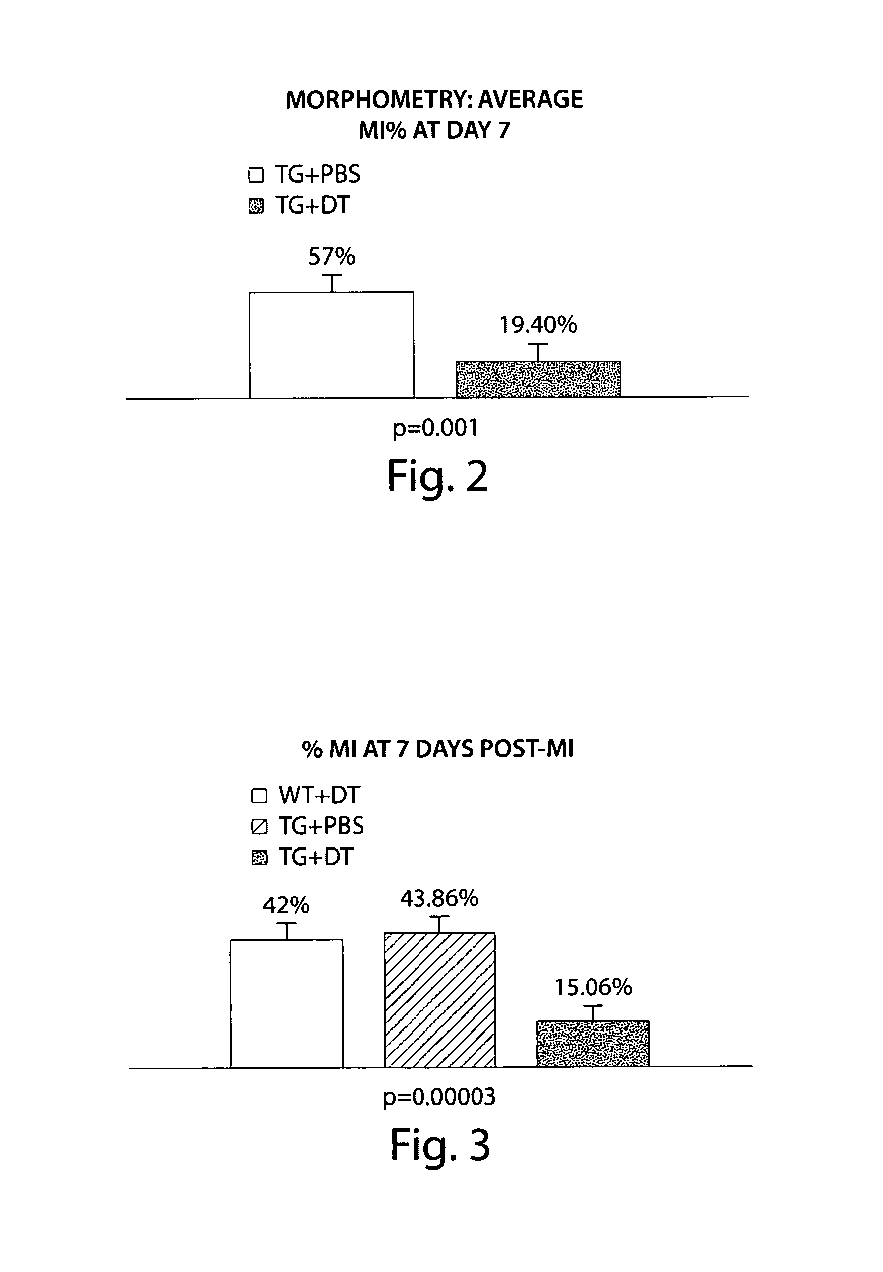
[0169]A single injection of DT has previously been shown to deplete DC with a nadir one to two days after injection and full recovery of DC number at seven days after injection. Jung et al. (2002) Immunity. 17:211-220. Transgenic DTR mice and wild type mice were randomized to injection of DT during surgery for induction of MI and physiologic readouts were acquired after MI at the time point when DC were most abundant in the MI area, i.e., seven days after MI (FIG. 1). Transgenic mice injected with phosphate buffered saline (PBS) served as controls for DT injection.
[0170]At seven days after MI there was a significant reduction in MI size by morphometry (FIG. 2) as well as by echocardiography (FIG. 3). There was also significantly greater preservation of LV function in those animals that had received DT (FIG. 4).
PUM
| Property | Measurement | Unit |
|---|---|---|
| time | aaaaa | aaaaa |
| diameter | aaaaa | aaaaa |
| molecular mass | aaaaa | aaaaa |
Abstract
Description
Claims
Application Information
 - R&D
- Intellectual Property
- Life Sciences
- Materials
- Tech Scout
- Unparalleled Data Quality
- Higher Quality Content
- 60% Fewer Hallucinations
Browse by: Latest US Patents, China's latest patents, Technical Efficacy Thesaurus, Application Domain, Technology Topic, Popular Technical Reports.
© 2025 PatSnap. All rights reserved.Legal|Privacy policy|Modern Slavery Act Transparency Statement|Sitemap|About US| Contact US: help@patsnap.com


