Cold organ preservation composition and method of use
a technology of organs and compositions, applied in the field of cold organ preservation compositions, can solve the problems of increasing immunogenicity, affecting the survival of animals,
- Summary
- Abstract
- Description
- Claims
- Application Information
AI Technical Summary
Benefits of technology
Problems solved by technology
Method used
Image
Examples
example 1
Preparation of Cold Organ Preservation Compositions
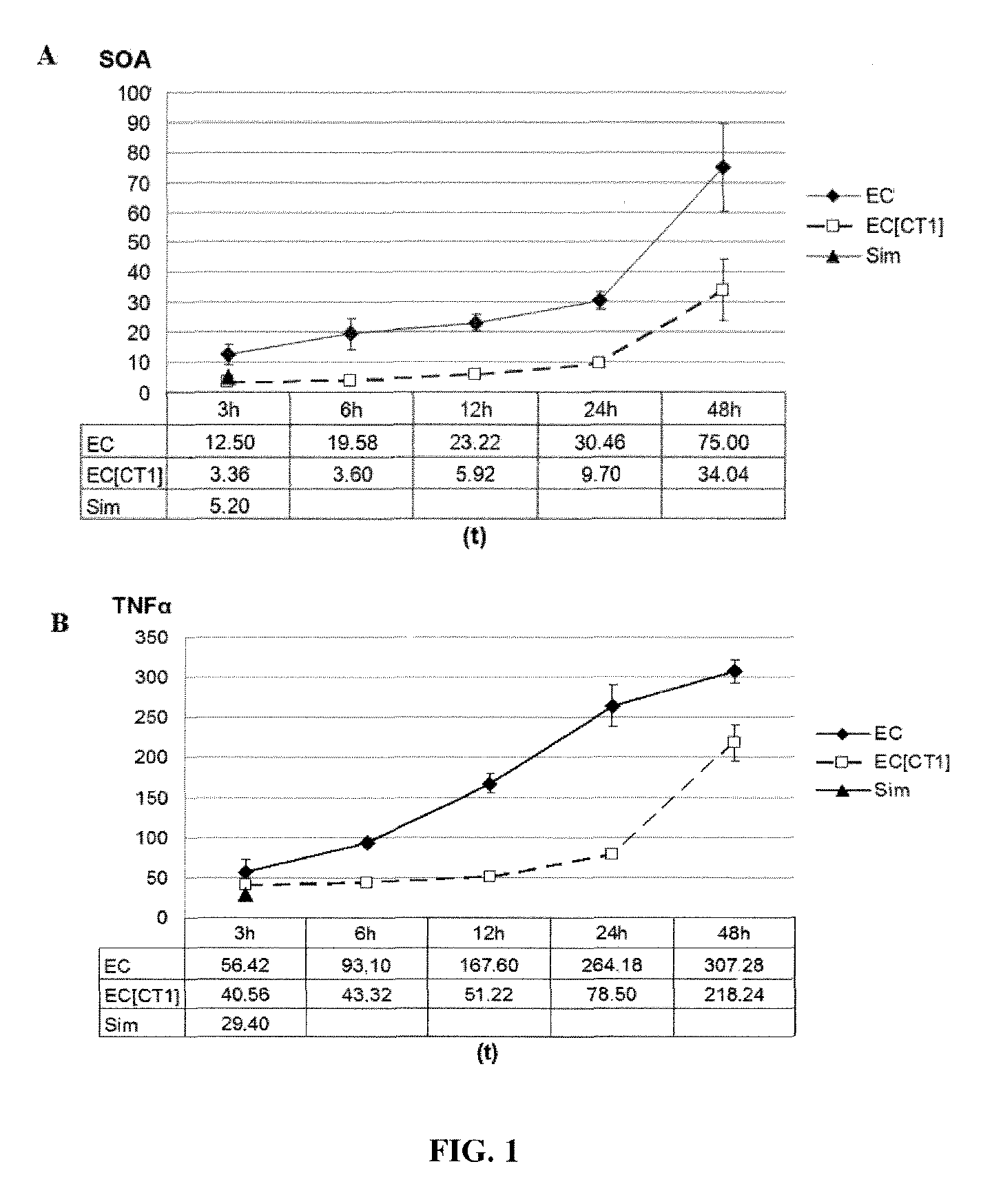
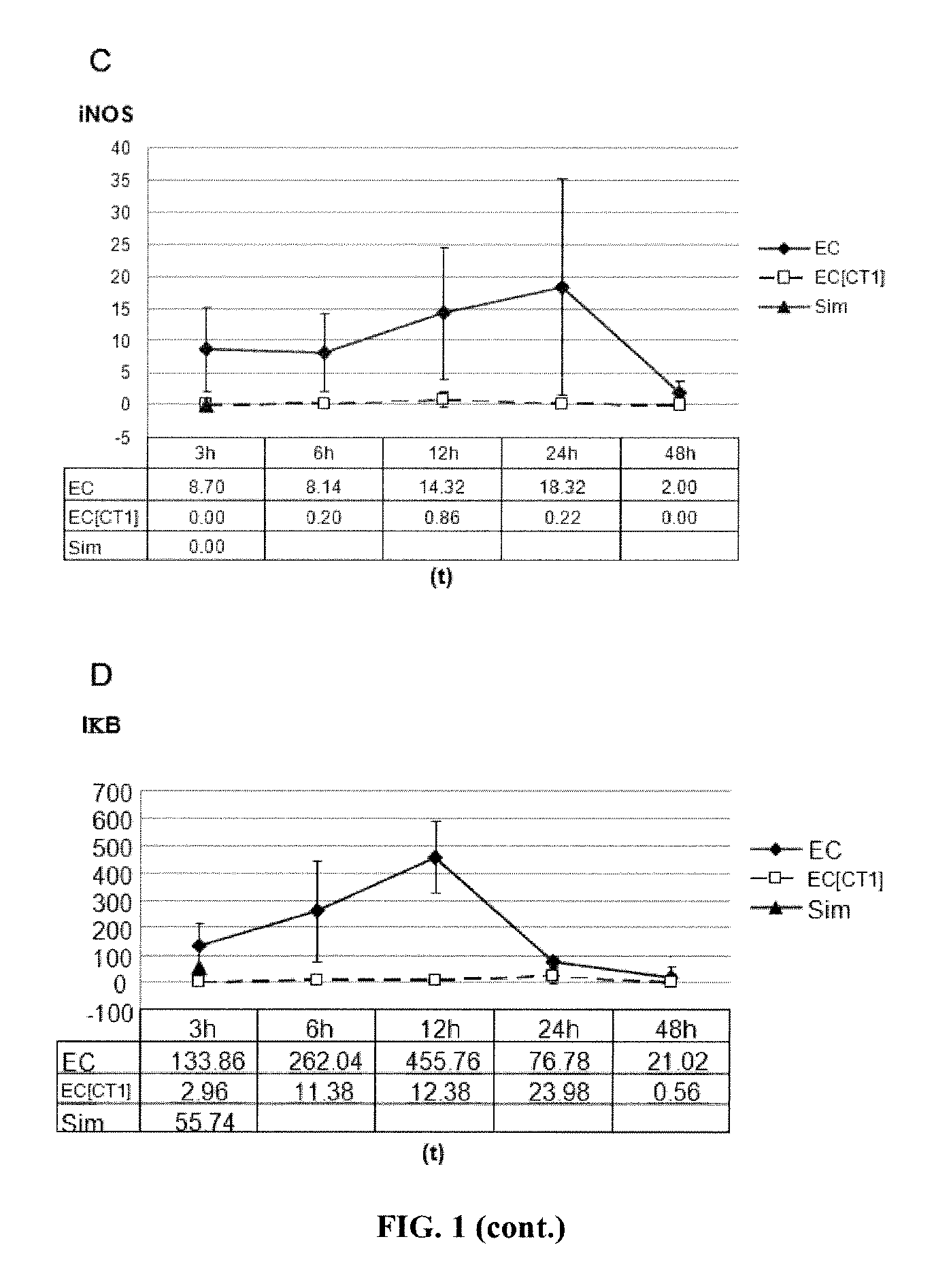
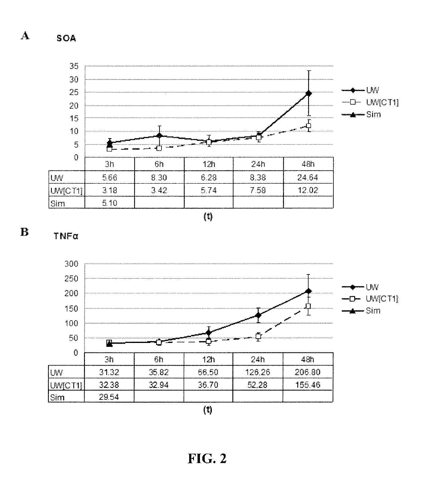
[0200]Different cold organ preservation compositions were prepared, supplemented with cardiotrophin-1 (CT-1), using the Euro-Collins solution (EC) or, alternatively, the University of Wisconsin solution (UW), as the base cold organ preservation solution.
[0201]Since the cold preservation solutions were to be assayed in experimental rat models, in order to supplement the media, rat CT-1 supplied by DRO Biosystems was used. It was produced in E. Coli BL21 (Invitrogen, Barcelona, Spain) as a fusion protein with an amino-terminal histidine tail. The expression of the recombinant protein was induced with IPTG (Sigma) for a final concentration of 0.5 mM in 2 hours at 25° C. After homogenising the bacteria pellet, the His-CT-1 fusion protein was purified by means of nickel affinity column chromatography (Qiagen, Barcelona, Spain). The sequence of CT-1 used is SEQ ID NO: 2:
[0202]
MRGSHHHHHH GMASMTGGQQ MGRDLYDDDD KDRWGSMSQR EGSLEDHQTD SRFSFLPH...
example 2
Studies of Cold Renal Preservation With a Cardiotrophin-1 Concentration of 0.1 mg / l
[0207]Wistar rats were used (225-300 g / rat males for the first study, and 225-250 g / rat males for the second study). The rats were anaesthesised by intraperitoneal injection with ketamine chloride (75 mg / kg)+diazepam (50 mg / kg) and atropine (20 mg / kg). A median laparotomy was performed and the retroperitoneum was dissected in order to expose the abdominal aorta and the renal arteries to the aortic bifurcation (iliac arteries). Subsequently, the renal vessels were dissected by means of ligation of the inferior and gonadal suprarrenal branches; and the ureter was dessicated at the proximal half, respecting the periurethral fat.
[0208]Subsequently, for perfusion of the organ (washing perfusion), the aorta was clamped below the renal artery exit, thereby maintaining the vascularisation thereof. The aortic catheterisation was performed by means of transverse aortotomy on the front face, following distal lig...
example 3
Study of Cold Renal Preservation With a Cardiotrophin-1 Concentration of 0.2 mg / l
[0218]Male Wistar rats, 225-250 g / rat, were used. Prior to the extraction, a cold perfusion of the kidney and the liver was performed with the preservation composition at 4° C. Subsequently, during the same surgical action, the two organs were separately extracted. In the first place, the ligaments of the liver were sectioned. Subsequently, the bile duct was sectioned in order to simulate the transplantation donour surgery, and the right renal vein and the right renal artery were ligated. Subsequently, a double ligation of the two ends of the pyloric vein was performed, and it was dessicated. Thereafter, 1 ml of heparinised saline solution was administered by intravenous injection. The aorta was cannulated with a 20G catheter and perfused at a rate of 4 ml / min with 12 ml of the cold preservation composition. Immediately following the perfusion, the inferior vena cava was dissected in order to allow for ...
PUM
 Abstract
Description
Claims
Application Information
 - R&D
- Intellectual Property
- Life Sciences
- Materials
- Tech Scout
- Unparalleled Data Quality
- Higher Quality Content
- 60% Fewer Hallucinations
Browse by: Latest US Patents, China's latest patents, Technical Efficacy Thesaurus, Application Domain, Technology Topic, Popular Technical Reports.
© 2025 PatSnap. All rights reserved.Legal|Privacy policy|Modern Slavery Act Transparency Statement|Sitemap|About US| Contact US: help@patsnap.com


