Method for identifying antigen-specific regulatory T cells
a technology of antigen-specific regulatory cells and t cells, which is applied in the field of methods for identifying and isolating antigen-specific regulatory t cells, can solve the problems of unable to isolate viable treg populations for functional studies or ex vivo expansion as a prelude to therapeutic administration, and the accuracy of identifying viable treg using cd4, cd25 and foxp3 markers is problemati
- Summary
- Abstract
- Description
- Claims
- Application Information
AI Technical Summary
Benefits of technology
Problems solved by technology
Method used
Image
Examples
example 1
Antigen Stimulation and Cell Identification and Isolation
[0134]Materials and Methods
[0135]Reagents
[0136]The antigens used were cytomegalovirus (CMV) lysate (22), mycobacterial antigen lysates (Mycobacterial tuberculosis (MTB) or Mycobacterial Avium intracellular complex (MAI), CSL, Melbourne, Australia), CMV-peptide (P1) (23, 24). Staphylococcal enteroantigen B (SEB) (Sigma-Aldrich Co., St Louis, Mo., United States of America) was used as a mitogen.
[0137]The monoclonal antibodies (mAbs) used were anti-CD3-PerCP-Cy5.5, anti-CD4-PE-Cy7, anti-CD45RO-FITC, anti-CD25-APC, anti-CD134-FITC, anti-CD134-PE and anti-Foxp3 (clone 259D) (all from Becton-Dickinson, San Jose, Calif., United States of America); anti-CD45RO-ECD (Beckman Coulter, Hialeah, Fla., United States of America); CD127-PE (Immunotech, Marseille, France); anti-CD127-Pacific Blue and anti-Foxp3-APC (clone PCH101) (eBiosciences, San Diego, Calif., United States of America); and CD39-PE (Serotec, Oxford, UK). All antibodies were...
example 2
Identification of CD4+CD25+CD134+Foxp3+ Antigen-specific Treg from Whole Blood
[0142]Foxp3 is an accepted marker of Treg. It was investigated whether antigen-specific CD4+ Treg could be identified in whole blood by detecting the co-expression of CD4, CD134, CD25 and Foxp3 following in vitro stimulation with antigen
[0143]Materials and Methods
[0144]Whole blood from two healthy controls (donor 1 and donor 2), who were known to have active immune responses to MAI, MTB or CMV was stimulated in vitro with no antigen, CMV, MTB or MAI lysates as described in Example 1. In a second experiment, whole blood from donor 1 was stimulated with no antigen, CMV-P1 antigen or SEB mitogen (as a positive control) for 24 to 40 hours as described in Example 1.
[0145]The cells were then stained with the combination of mAbs consisting of anti-CD3, anti-CD4, anti-CD25, anti-CD134 and anti-Foxp3 antibodies. Cell staining was analysed on a three-laser LSR II flow cytometer (Becton-Dickinson). Antigen-specific T...
example 3
Isolation of CD4+CD25+CD39+CD134+ Viable Antigen-Specific Treg from Whole Blood
[0152]Detection of Foxp3 requires cell permeabilisation, which is problematical when it is desirable to have live cells following cell staining. CD39 has recently been reported to be a useful alternative marker for detecting Treg. In this example, the present applicant investigated whether antigen-specific CD4+Treg could be identified in whole blood following in vitro stimulation with antigen by detecting the co-expression of CD4, CD25, CD134 and CD39.
[0153]Materials and Methods
[0154]Whole blood from donor 1 was stimulated with no antigen, CMV-P1 antigen or SEB mitogen (as a positive control) for 24 hr as described in Examples 1 and 2. The cells were then stained with the monoclonal antibodies consisting of anti-CD3, anti-CD4, anti-CD39, anti-CD25, anti-CD134 and anti-Foxp3 as described in Examples 1 and 2. CD3+CD4+ cells were gated.
[0155]Results
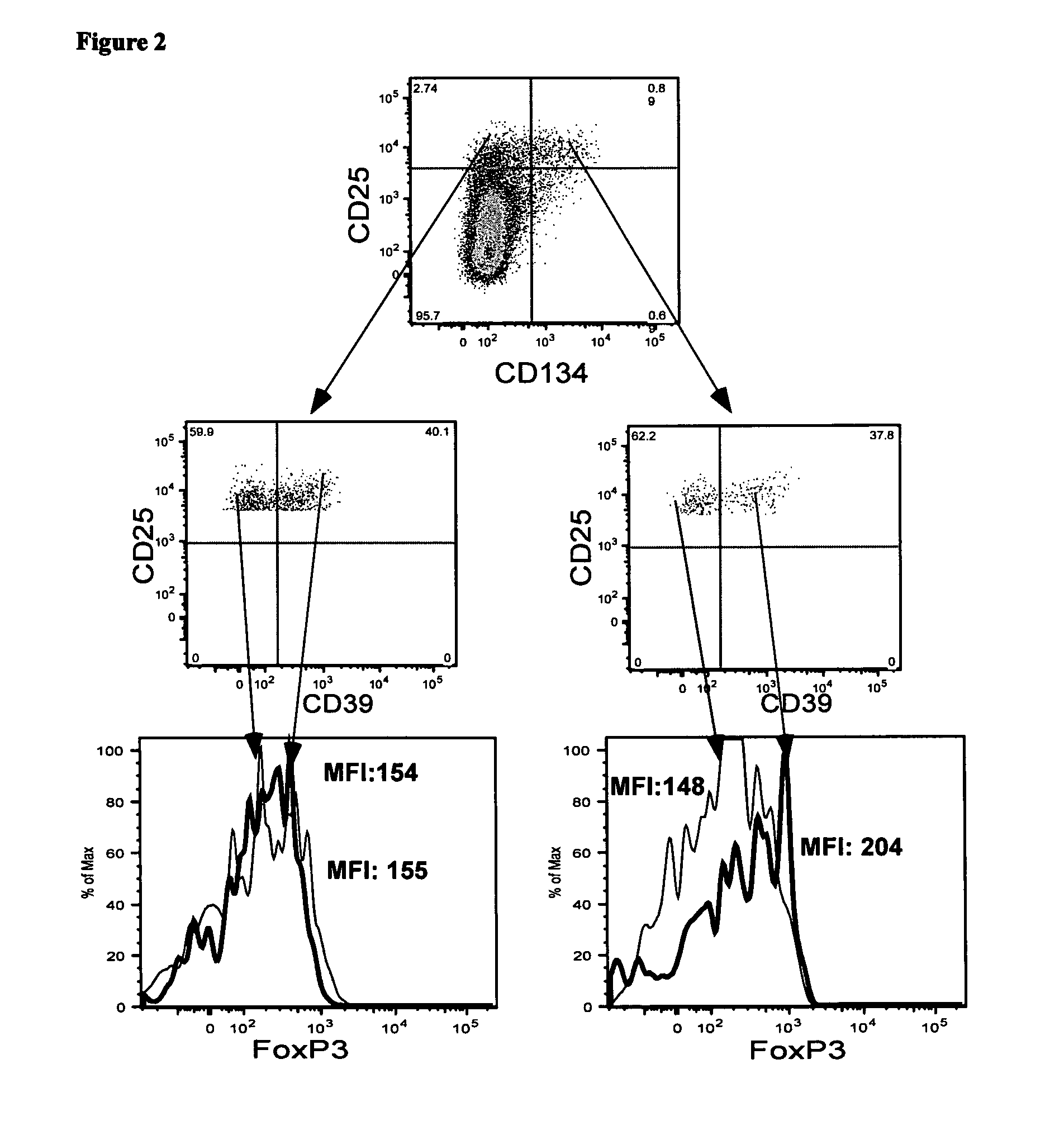
[0156]The plots shown in FIG. 2 demonstrate that approximate...
PUM
| Property | Measurement | Unit |
|---|---|---|
| period of time | aaaaa | aaaaa |
| period of time | aaaaa | aaaaa |
| time | aaaaa | aaaaa |
Abstract
Description
Claims
Application Information
 - R&D
- Intellectual Property
- Life Sciences
- Materials
- Tech Scout
- Unparalleled Data Quality
- Higher Quality Content
- 60% Fewer Hallucinations
Browse by: Latest US Patents, China's latest patents, Technical Efficacy Thesaurus, Application Domain, Technology Topic, Popular Technical Reports.
© 2025 PatSnap. All rights reserved.Legal|Privacy policy|Modern Slavery Act Transparency Statement|Sitemap|About US| Contact US: help@patsnap.com


