Method for diagnosing melanocytic proliferations
a melanocytic and proliferation technology, applied in the field of melanocytic proliferation diagnosis, can solve problems such as characteristic staining sac pattern, and achieve the effect of diagnosing melanocytic proliferation
- Summary
- Abstract
- Description
- Claims
- Application Information
AI Technical Summary
Benefits of technology
Problems solved by technology
Method used
Image
Examples
example 1
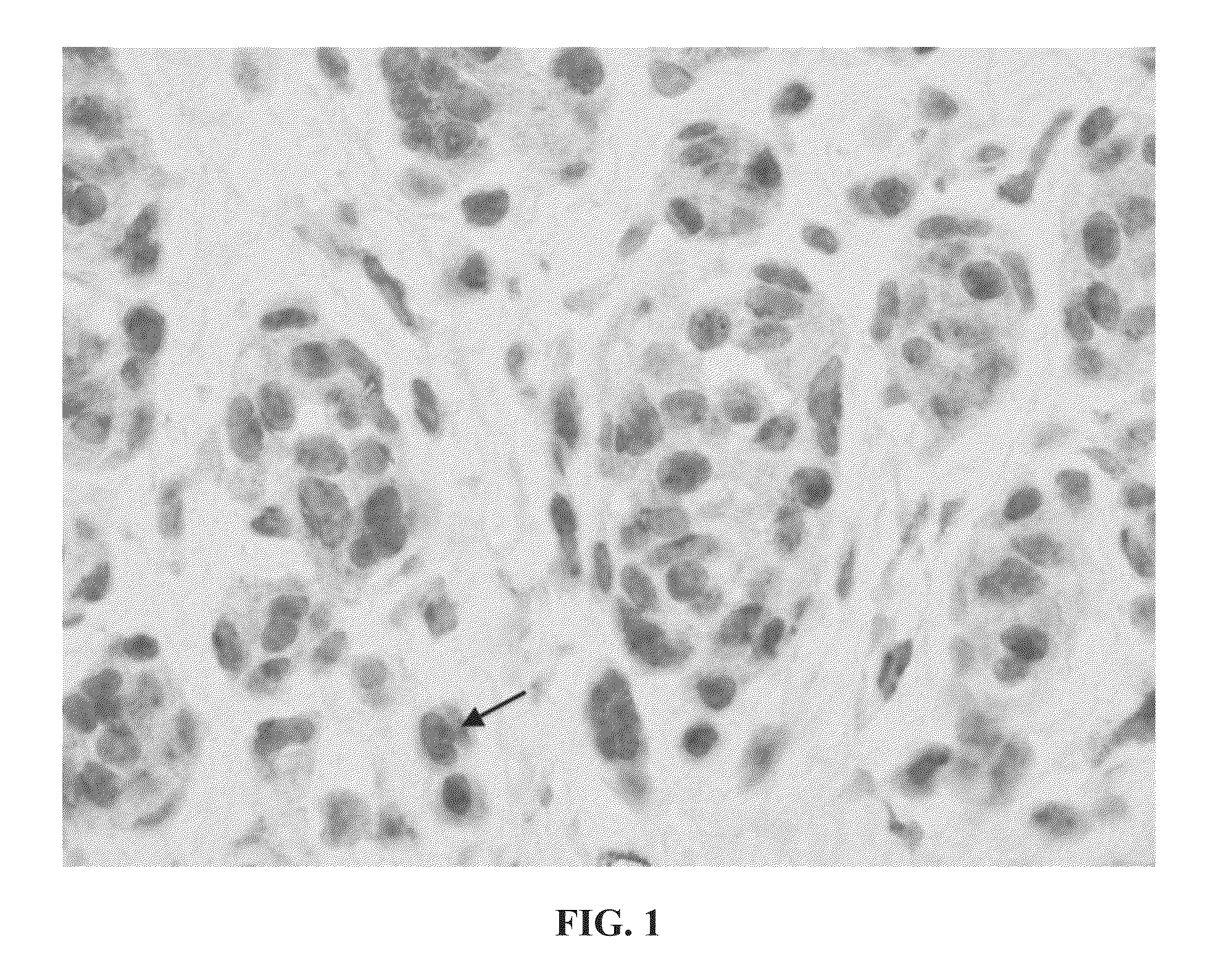
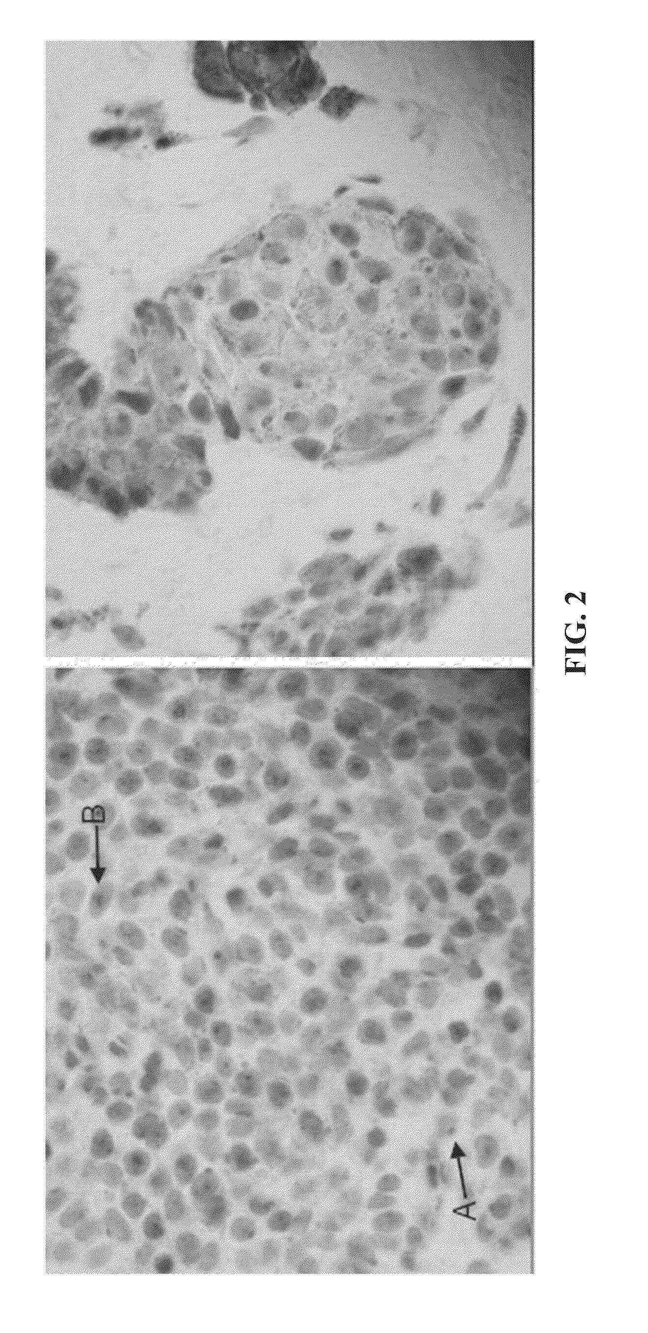
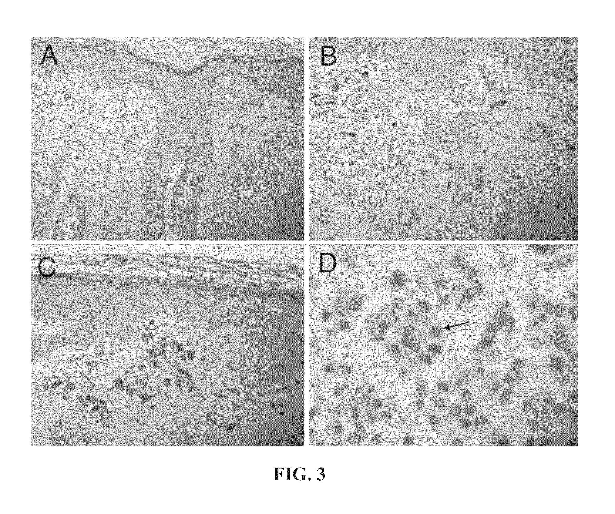
[0110]This example demonstrates the staining of a tissue sample with R21 antibody against sAC to provide an sAC staining pattern. All steps were performed using the Leica Microsystems BOND-MAX™ autostainer (Bannockburn, Ill.).
[0111]Dewaxing: Formalin-fixed, paraffin embedded samples of lesional melanocytes were baked at 60° C. for 30 minutes. Slides were then treated with a Leica Microsystems BOND DEWAX SOLUTION™ (AR9222) for 3 minutes at 72° C., followed by washes in BOND DEWAX SOLUTION™ first at 72° C. and then at ambient temperature. Finally, slides were washed three times with ethyl alcohol 200-proof (Pharmco-Aaper, Brookfield, Conn., 111000200) and three times with Leica Microsystems BOND WASH SOLUTION™ (AR9590).
[0112]Pre-treatment and blocking: Following the dewaxing procedure, the sections were pre-treated by two washes in Leica Microsystems BOND EPITOPE RETRIEVAL SOLUTION 1™ (AR9961), followed by BOND EPITOPE RETRIEVAL SOLUTION 1™ pre-treatment for 30 minutes at 100° C., and...
example 2
[0116]This example demonstrates that the analysis of a sAC staining pattern of a sample of lesional melanocytes from a subject can be used to provide a diagnosis of melanocytic proliferations for the subject.
[0117]Samples of lesional melanocytes were obtained from 140 different subjects and subjected to the procedures described in Example 1 to obtain or discern sAC staining patterns. The 140 sAC staining patterns were analyzed and grouped into categories pertaining to different diagnoses of melanocytic proliferations. The results of this analysis provided the following sAC staining pattern characteristics for each of the diagnoses of melanocytic proliferations.[0118](1) Benign nevus and capsular nevus of lymph nodes[0119](a) Dot-like Golgi staining in >50% of lesional melanocytes[0120](b) Weak nucleolar and incomplete granular nuclear staining in 50% of lesional melanocytes, on average[0121](c) Pan nuclear staining in [0122](2) Atypical nevus of special sites[0123](a) Pan-nuclear st...
example 3
[0178]This example compares the dot-like Golgi and pan-nuclear staining patterns in dysplastic nevi to the dot-like Golgi and pan-nuclear staining patterns in melanoma.
[0179]Loss of dot-like Golgi staining with gain of pan-nuclear staining appears to be indicative of melanoma. In the following table (Table 1), loss of dot-like Golgi staining (DG) is assumed when 25% total melanocytes have this staining pattern (range for nevi 0-25%; range for melanomas 50-90%). Melanomas were compared to a combined group of all moderately and severely dyspalstic nevi, with “all melanomas” representing superficial spreading, acral lentiginous, nodular, and lentigo maligna melanomas, and “SSM” representing superficial spreading melanomas.
[0180]To determine if particular staining patterns were present or absent in a given type of lesion more often than simple chance, a binomial proportion test was performed (50% assumption being simple chance) with a two-sided student t-test. To determine if there was ...
PUM
| Property | Measurement | Unit |
|---|---|---|
| size | aaaaa | aaaaa |
| temperature | aaaaa | aaaaa |
| temperature | aaaaa | aaaaa |
Abstract
Description
Claims
Application Information
 - Generate Ideas
- Intellectual Property
- Life Sciences
- Materials
- Tech Scout
- Unparalleled Data Quality
- Higher Quality Content
- 60% Fewer Hallucinations
Browse by: Latest US Patents, China's latest patents, Technical Efficacy Thesaurus, Application Domain, Technology Topic, Popular Technical Reports.
© 2025 PatSnap. All rights reserved.Legal|Privacy policy|Modern Slavery Act Transparency Statement|Sitemap|About US| Contact US: help@patsnap.com


