


Quick Research
Generate reliable direction feasibility study reports for your R&D in just a few steps.

Technical Q&A
Discover and master advanced knowledge NOW. Basics, ideas, possibilities, all at once.

Find Solutions
As an expert in R&D theories, this can generate solutions to your technical problems instantly.

Evaluate Feasibility
Analyze your overall solution with one click, know your potential R&D risks in advance.

Monitor Landscape
Get weekly tech updates, stay abreast of the latest tech innovations and key insights.
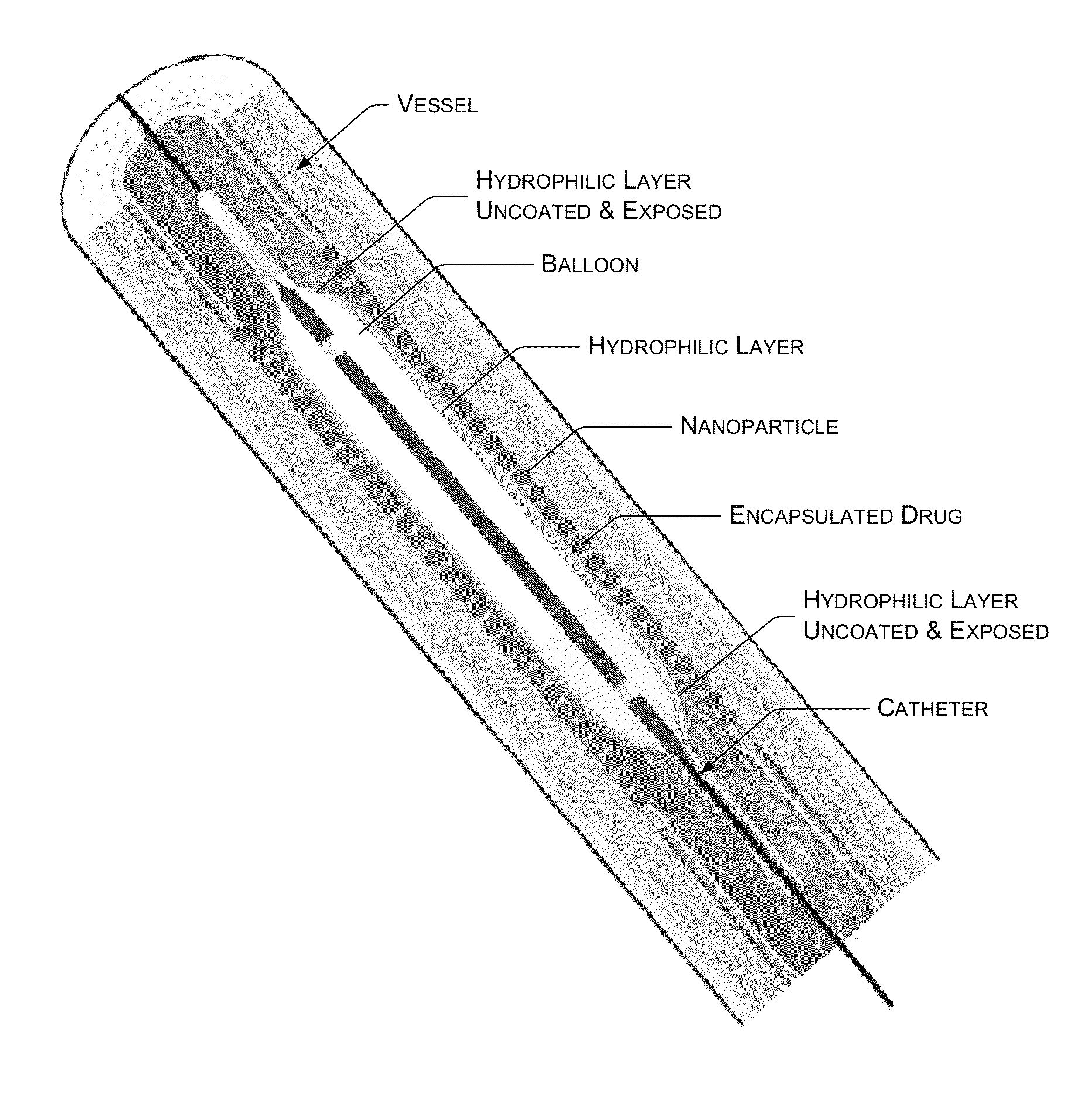
Balloon catheter providing decreased irritation and improved drug release and drug penetration
a balloon and balloon technology, applied in the field of medical devices, can solve the problems of ineffective penetration of micro-sized particles of drugs into tissues inflammation at the target site, and causing unwanted side effects
- Summary
- Abstract
- Description
- Claims
- Application Information
AI Technical Summary
Benefits of technology
Problems solved by technology
Method used
Image

Examples
example 1
[0057]Soya phospholipid was obtained from Lioid GMBH, Batch No.: 776114-1 / 906. Sirolimus was obtained from Fujan Chemicals, China with purity greater than 99.5%. The water, other solvents and reagents used were of HPLC grade. A polyamide catheter system with COPAN Co-Polyamide angioplasty balloon (herein after “the balloon system”) coated with Hydraflow® Hydrophilic coating (hereinafter “the hydrophilic surface”) was obtained from Minvasys, Paris, France.
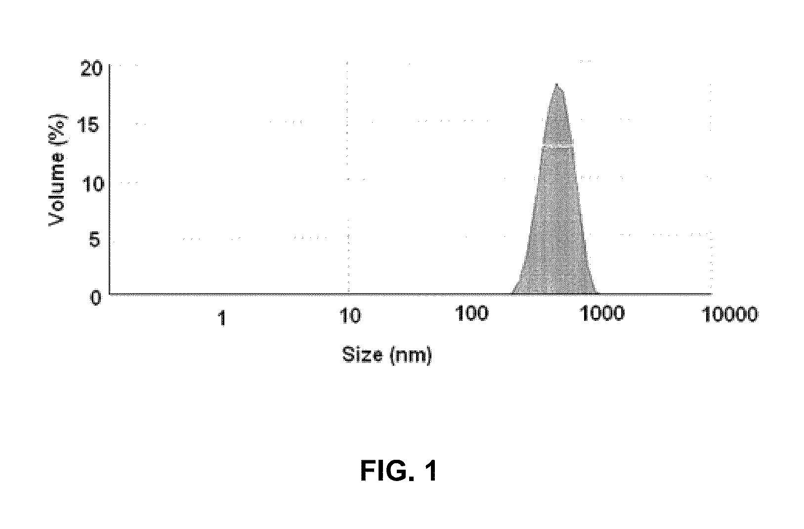
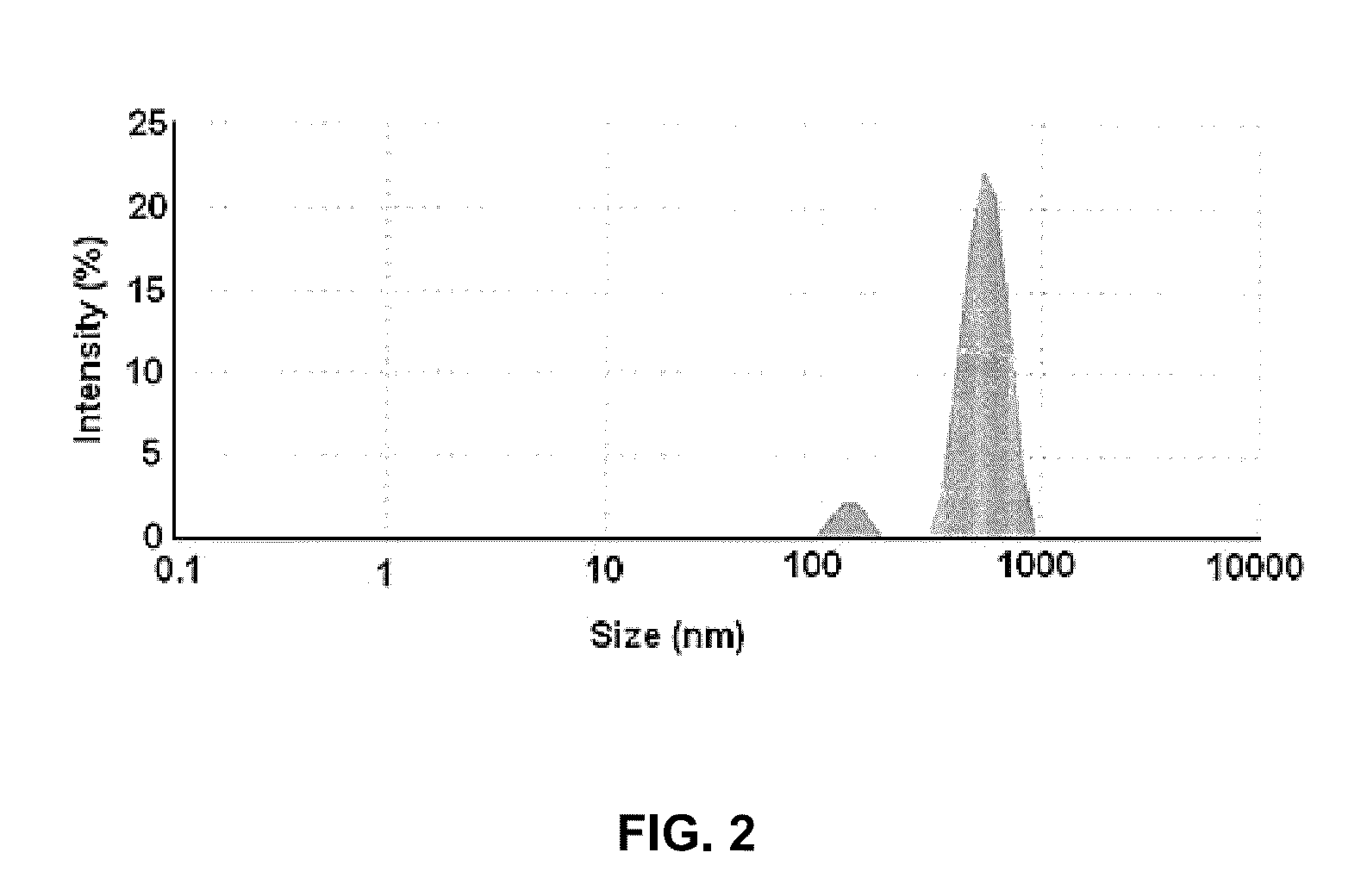
[0058]Soya phospholipid (20 mg w / w) was added to HPLC grade water (20 ml) followed by Tween 80 (5 mg) to obtain aqueous solution of soya phospholipid. The aqueous solution of soya phospholipid (20 ml) was subjected to a high speed homogenization at 15000-20000 rpm for 20 to 25 minutes in ice-cold water bath to obtain Solution A1. The Solution A1 thus obtained contained nano-particles of soya phospholipid. The solution A1 was subsequently analyzed for particle size detection using Malvern ZS90 (Malvern, UK) size detector. FIG. 1 illu...
example 2
[0063]The coated balloon was further evaluated in-vivo in 17 male New Zealand rabbits, 5 to 6 months old and weighing between 3 and 4 Kg (hereinafter “animals”) for PharmacoKinetic (PK) study and histological evaluation or Light Microscopy (LM). FIG. 3 is a table illustrating numbers assigned to 17 animals, stent type and location of stent in each animal and the type of study (PK / LM) conducted on the animals. Out of the 17 animals 9 (Animals 66 to 74) were used for PK study and 8 (Animals 75 to 82) were used for histological evaluation.
[0064]The coated balloons were inserted into both the iliofemoral arteries of the animals used for the PK study. The coated balloons were inflated twice in the iliofemoral arteries. The coated balloons were first inflated for 70 seconds at 7 ATM and then deflated. Again, the coated balloons were inflated for the second time for 60 seconds at 7 ATM and then deflated and withdrawn.
[0065]A pre-mounted stent (3.0 mm×12-14 mm) i.e. a stent mounted on the c...
example 3
[0072]6 Brazilian pigs (Hereinafter “the animals”) weighing about 25 to 30 kg were selected for the study. Bare metal stent (Cronus®, obtained from, Scitech, Brazil), with sizes of stent ranging from 2.5*13 mm to 3.0*13 mm and the coated balloons with sizes of about 3.0*15 mm were used. The stents were deployed in three vessels i.e. LAD (Left Interior Descending), LCX (Left Circumflex) and RCA (Right Coronary Artery of each animal by using: a) the coated balloon (Sirolimus), b) the coated balloon (Paclitaxel) and c) a bare balloon. The Paclitaxel coated balloon was prepared by replacing sirolimus in example 1 with Paclitaxel and rest of the process was kept same. The balloon to artery ratio for each coated balloon was approximately 1.1:1.0 to 1.2:1.0. Each of the coated balloon and the bare balloon were inflated for 60 seconds. The animals with sirolimus coated balloon were labeled “Sirolimus”, the animals with Paclitaxel coated balloon were labeled “Paclitaxel” and the animals with...
PUM
| Property | Measurement | Unit |
|---|---|---|
| average diameter | aaaaa | aaaaa |
| diameter | aaaaa | aaaaa |
| diameter | aaaaa | aaaaa |
Abstract
Description
Claims
Application Information
 - R&D Engineer
- R&D Manager
- IP Professional
- Industry Leading Data Capabilities
- Powerful AI technology
- Patent DNA Extraction
Browse by: Latest US Patents, China's latest patents, Technical Efficacy Thesaurus, Application Domain, Technology Topic, Popular Technical Reports.
© 2024 PatSnap. All rights reserved.Legal|Privacy policy|Modern Slavery Act Transparency Statement|Sitemap|About US| Contact US: help@patsnap.com