
Biosensor and use thereof to identify therapeutic drug molecules and molecules binding orphan receptors

a biosensor and drug candidate technology, applied in the field of recombinant dna technology, can solve the problems of insufficient information about the behavior of these signaling pathways, undesired serious limitation in the methods available to screen drug candidates non-invasively using mammalian g protein coupled receptors and g proteins, lack of information about temporal changes and spatial localization, etc., to achieve the effect of increasing the range and depth of candidate molecules

Inactive Publication Date: 2013-11-12
WASHINGTON UNIV IN SAINT LOUIS
View PDF3 Cites 0 Cited by
  • Summary
  • Abstract
  • Description
  • Claims
  • Application Information

AI Technical Summary

Benefits of technology

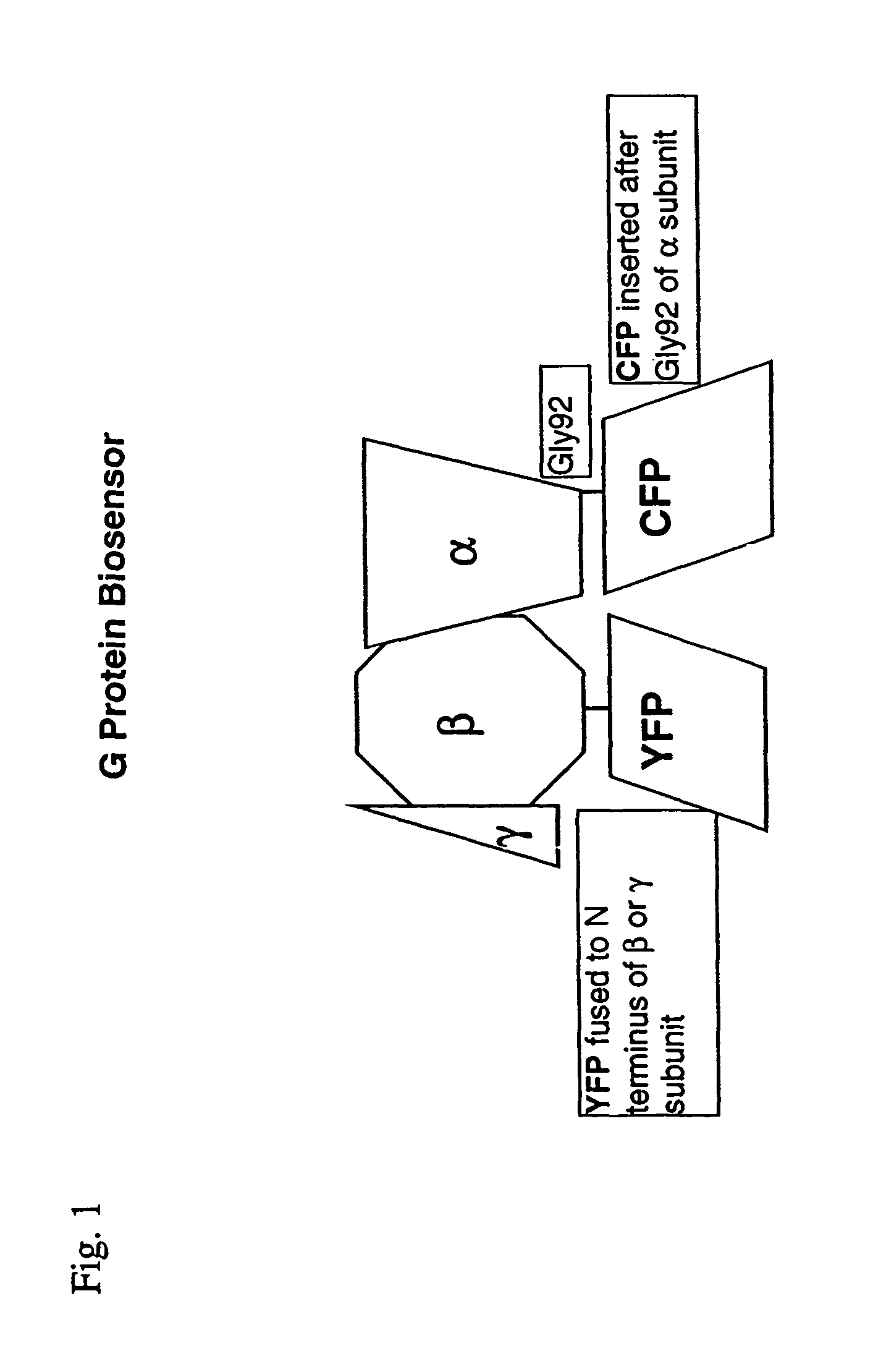
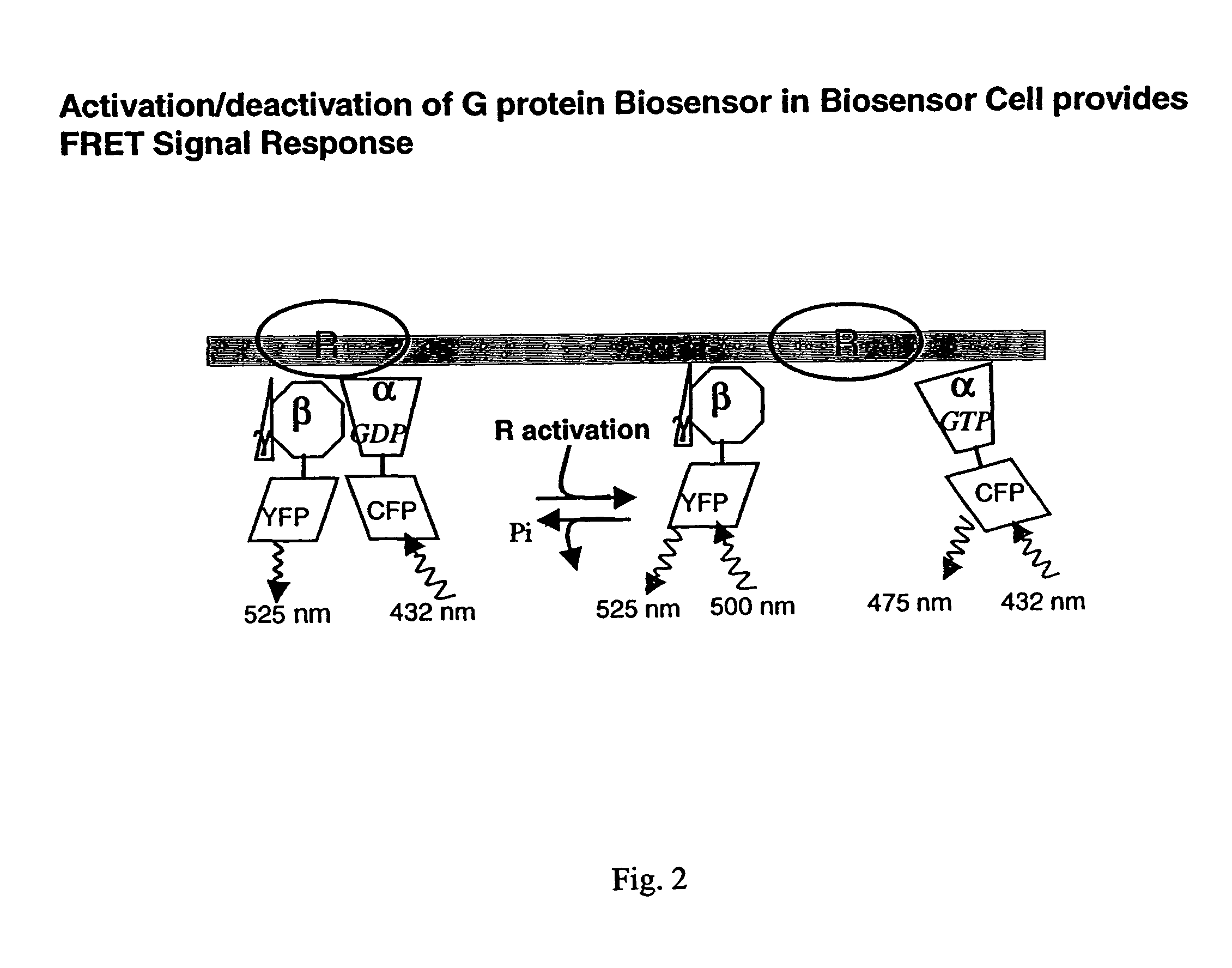
The patent describes a method for creating a live functional biosensor that can detect and measure the activity of G proteins in cells. This biosensor consists of a fusion protein containing a fluorescent protein and a G protein subunit, which can interact with each other to produce a fluorescence signal. The biosensor can be used to screen for natural or synthetic molecules that can activate or block the receptors that G proteins are associated with. The method is non-invasive and can provide information about the activity of G proteins in real-time and in their natural environment. The patent also describes a method for identifying and classifying therapeutic molecules based on their activity at different receptors.

Problems solved by technology

However, G protein signaling operations occur in intact living cells subject to constraints of dynamic equilibrium, which are disrupted when cells are broken.
Unfortunately, relatively little information is at present available about the behavior of these signaling pathways in an intact living mammalian cell because methods have not been available for their observation.
While the three dimensional structure of the G protein coupled receptor and newer methods of rational drug design increase the range and depth of candidate molecules are available, there is at present an undesired serious limitation in methods available to screen drug candidates non-invasively using mammalian G protein coupled receptors and G proteins.
There is also a lack of information about the temporal changes and spatial localization of the effects of candidate therapeutic molecules in an intact living cell.

Method used


Image

  • Biosensor and use thereof to identify therapeutic drug molecules and molecules binding orphan receptors
  • Biosensor and use thereof to identify therapeutic drug molecules and molecules binding orphan receptors
  • Biosensor and use thereof to identify therapeutic drug molecules and molecules binding orphan receptors

Examples


example 1

[0159]A mammalian G protein biosensor was prepared following the aforedescribed procedure herein functional and produced a FRET signal to test whether FRET occurred from α-CFP to β-YFP. YFP was bleached by illuminating for 4 min using the D500 / 20× excitation filter (YY set). The intensity of fluorescence emission from CFP was examined before and after bleaching. The CC signal increased about 3 to about 7% after YFP was bleached indicating that the biosensor cell was function and that FRET did occur from CFP to YFP in these fusion proteins This result also showed that the subunit fusions do form a heterotrimer.

[0160]The biosensor cell of this biological system is living because it has been cultured on the cover glass to which they are attached during operation and they have multiplied there and also respond to an extracellular signals with expected physiological response. The ability to reproduce and response to the environment characterizes them as living.

[0161]The biosensor cell is...

example 2

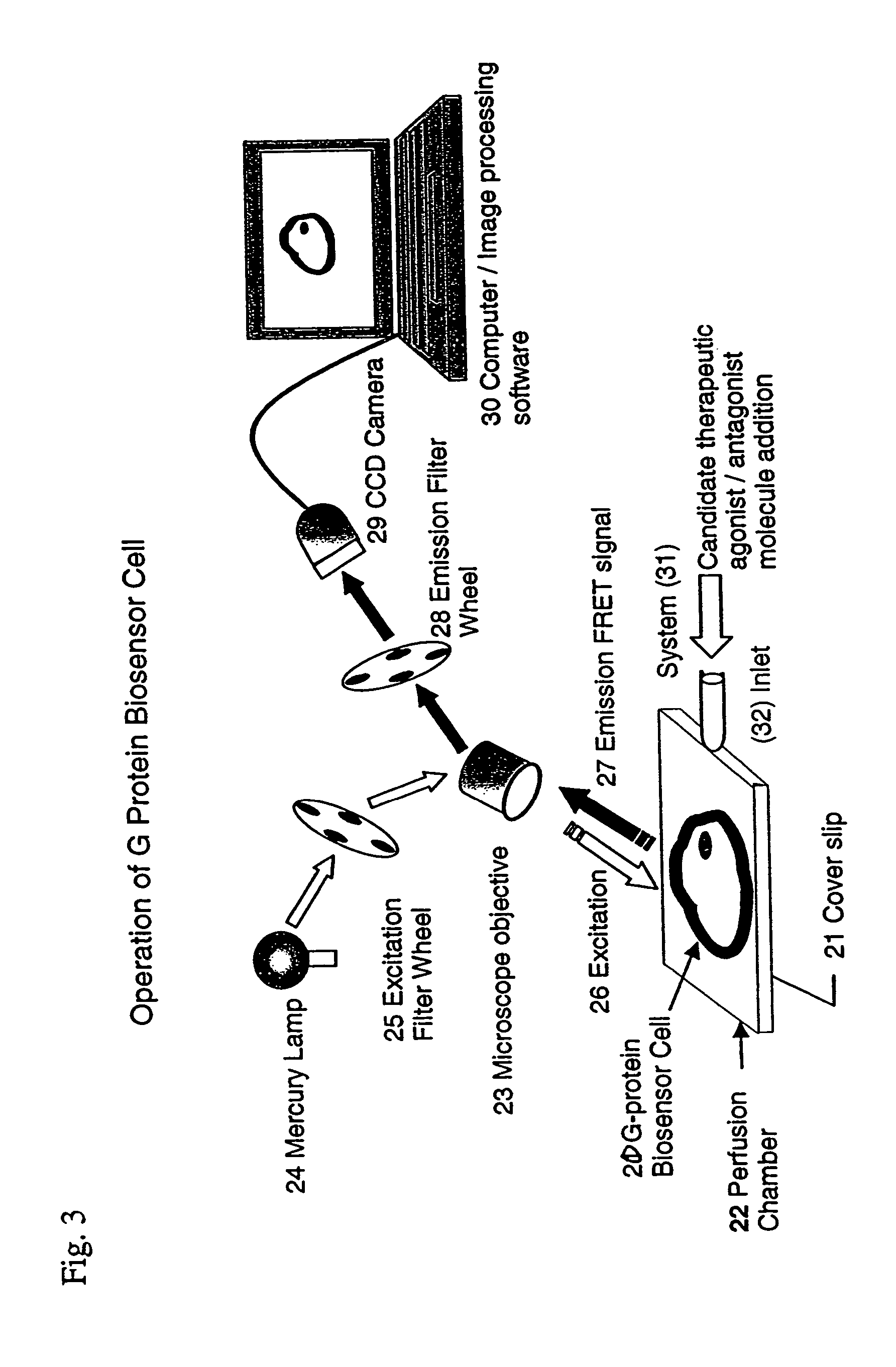
[0165]The functional G protein biosensor cell responded to an agonist molecule. Images of biosensor cells were acquired (captured) at regular time intervals of 15 sec before and after the addition of a muscarinic acetylcholine receptor agonist drug, carbachol. The FRET profile of the CY / CC ratio was plotted over time and is shown in FIG. 5. The FRET signal intensity decreases in the presence of the agonist drug. FRET images of biosensor cells were acquired and analyzed before and after exposure to carbachol as described in the Procedures for Design and Operation following. Timing of various agonist additions to the biosensor cell are indicated with arrows on FIG. 5. Plots show a downward trend because of partial bleaching over the period of the test. The graph is representative of data from ten tests. The results show that the ratio decreases strikingly when the muscarinic receptor agonist drug, carbachol is introduced into the perfusion chamber containing the cells.

example 3

[0166]The functioning G protein biosensor cell responded quantitatively and reproducibly to an agonist drug. The response of biosensor cells to the addition of varying concentrations of carbachol were measured as described earlier. The FRET signal intensity changes is directly and positively correlated with increases in agonist drug concentration (FIG. 6). Details are provided in Procedures for Design and Operation. Points are means±SEM of three tests. The data curve was fitted using GraphPad (Prizm). FRET signal intensity is decreased by agonist drug addition. Tests were performed as described hereinafter in Detailed Description of the Invention.

[0167]The EC50 for carbachol activation of the G protein is about 600 nM which is consistent with the EC50 for carbachol mediated M2 activation of a G protein measured in a reconstituted system (15).


PUM


Abstract

G protein biosensors comprising mammalian G protein subunits fused to fluorescent proteins emitting a FRET signal expressed in living intact functional cells. The intensity of the FRET signal is strongly responsive to the activation state of the biosensors. The biosensors respond reproducibly to agonist and antagonist drug molecules specific for G protein coupled receptors. The biosensors have utility in identifying and classifying candidate therapeutic drugs as to their therapeutic value.

Description

[0001]This application claims the individual and collective benefit of U.S. provisional patent application U.S. Ser. No. 60 / 445,113 filed Feb. 5, 2003 and U.S. provisional application U.S. Ser. No. 60 / 493,952 filed Aug. 8, 2003, both pending, each of which is incorporated herein in its respective entirety by reference.STATEMENT REGARDING FEDERALLY SPONSORED RESEARCH AND DEVELOPMENT[0002]This invention was made with government support under Grant Number GM46963 awarded by the National Institute of Health. The government has certain rights in the invention.FIELD OF THE INVENTION[0003]This invention relates to recombinant DNA technology and the preparation and operation of a functional biosensor capable of and capably operating in a living intact functional cell. More particularly, this invention relates to mammalian receptor stimulated G proteins and to a method of traditional or high content screening of candidate molecules non-invasively using a functional biosensor comprising G pro...

Claims


Application Information

Patent Timeline
IPC IPC(8): G01N33/53C12N5/07C12M1/34G01N33/50G01N33/542G01N33/58
CPCG01N33/5005G01N33/5008G01N33/5041G01N33/542G01N33/582G01N2333/4719
Inventor GAUTAM, NARASIMHANAZPIAZU, INAKI
Owner WASHINGTON UNIV IN SAINT LOUIS
Learn More