Cell culture apparatus and associated methods
a cell culture apparatus and cell technology, applied in the field of cell culture apparatus, can solve the problems of unable to grow into a 3-dimensional mass representative of in vivo tissue, and reducing the number of cells in the cell culture apparatus
- Summary
- Abstract
- Description
- Claims
- Application Information
AI Technical Summary
Benefits of technology
Problems solved by technology
Method used
Image
Examples
example embodiments
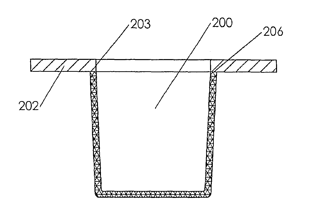
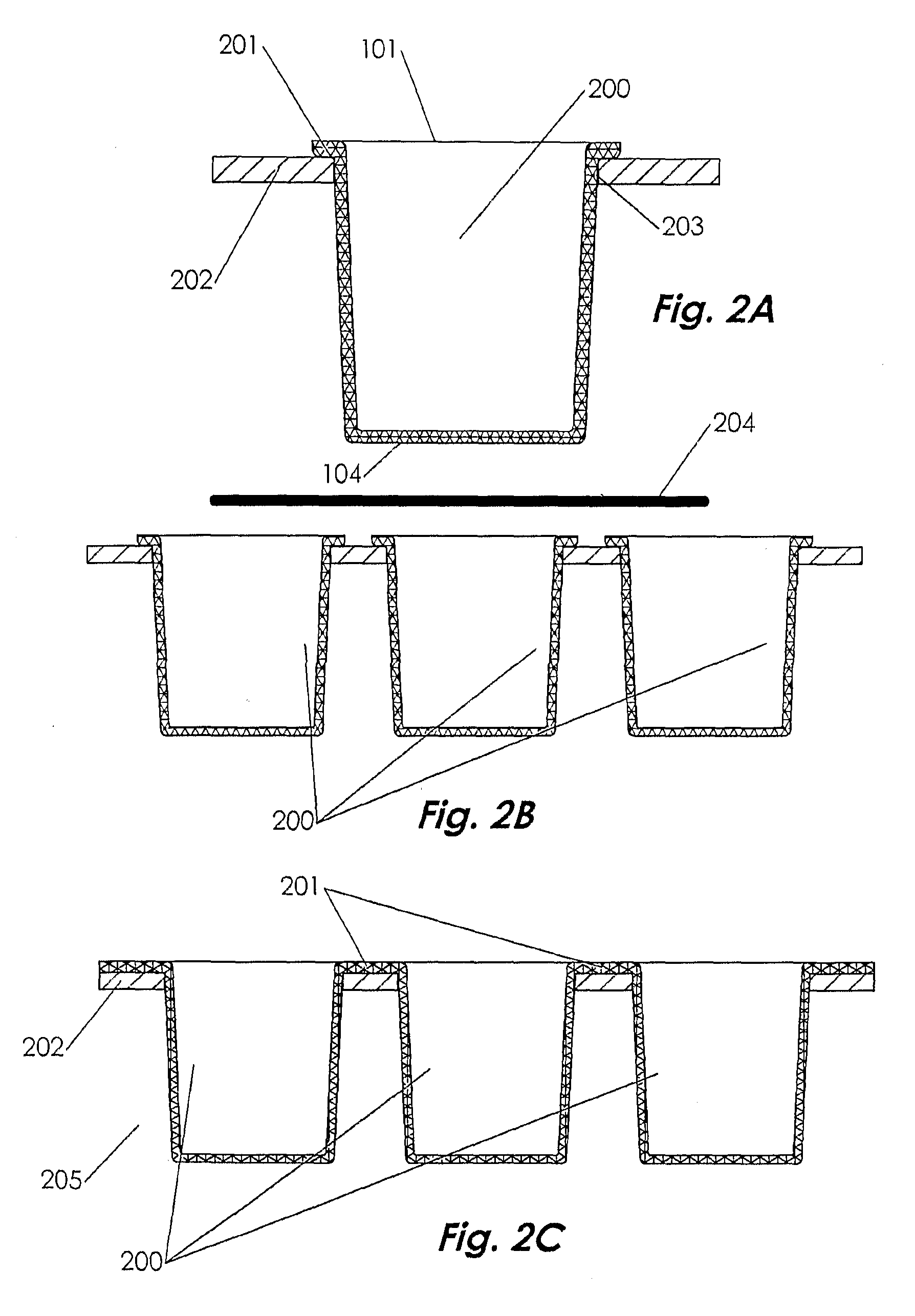
[0048]FIGS. 1A-1D show an embodiment of the cell culture apparatus in the basic form. The apparatus is composed of a vessel 100 or plurality of vessels in which each vessel comprises a bottom 101 and peripheral sides 102 with an open orifice at the top 110. The bottom is further comprised of an inner surface 103 and an outer surface 104; likewise, the sides also have an inner surface 105 and outer surface 106. The bottom and sides may be connected without transition radii; conversely, the bottom and sides may have an inner radius 107 connecting inner surfaces 103 and 105 and an outer radius 108 connecting outer surfaces 104 and 106. The bottom and sides are molded integrally and are composed of a gas-permeable material. Typically, the bottom and sides of the vessel are molded in a single action and as such are of the same gas permeable material. Alternately, the bottom and sides may be of different gas-permeable materials molded in a two-shot or overmold process. The bottom inner su...
example 1
Viable Multilayer Growth of Hep G2 Cells without Passaging
[0097]Hep G2 cells (human liver carcinoma) were cultured to approximately 12 cell layers deep for a period of over 180 days without passaging (note: In conventional TC-treated polystyrene labware these cells normally grow to monolayer confluence with various small multilayered foci; passaging is typically required every 3 to 4 days to avoid senescence / cell death). The cells were cultured in a vessel with an inside diameter of approximately 13.5 mm, an inside depth of approximately 17.0 mm and a wall thickness of approximately 0.75 mm, composed of compression-molded medical-grade silicone, Shore A40 durometer (Med-4940, NuSil Technologies, Carpinteria, Calif.). The mold surface which formed the inner bottom surface of the vessel was textured using 400 grit sandpaper, applied using a lathe. The mold surface which formed the outer bottom surface of the vessel was highly polished. After molding, the vessel was cleaned using a fiv...
example 2
Multilayer Growth of NIH 3T3 Cells without Passaging
[0099]NIH 3T3 cells (embryonic mouse fibroblast) were cultured into complex 3-dimensional structures for a period of over 4 months without passaging (note: In conventional TC-treated polystyrene labware these cells are normally highly contact inhibited and grow just to monolayer confluence prior to senescence; passaging is typically required every 3 to 4 days to avoid senescence). The vessel geometry and preparation is the same as Example 1. Similarly, the coating material and its application is the same as Example 1. NIH 3T3 cells were seeded at a density of 100,000 cells / vessel. An enhanced media formulation specific to this cell line is used and media is replaced and replenished on a 3-day cycle. After growing to confluence and then into multilayers, the cells commonly form large globular aggregates which can be seen with the naked eye. Fibrous cellular structures typically emanate from the globular aggregates, often connecting ...
PUM
| Property | Measurement | Unit |
|---|---|---|
| diameter | aaaaa | aaaaa |
| height | aaaaa | aaaaa |
| height | aaaaa | aaaaa |
Abstract
Description
Claims
Application Information
 - R&D
- Intellectual Property
- Life Sciences
- Materials
- Tech Scout
- Unparalleled Data Quality
- Higher Quality Content
- 60% Fewer Hallucinations
Browse by: Latest US Patents, China's latest patents, Technical Efficacy Thesaurus, Application Domain, Technology Topic, Popular Technical Reports.
© 2025 PatSnap. All rights reserved.Legal|Privacy policy|Modern Slavery Act Transparency Statement|Sitemap|About US| Contact US: help@patsnap.com


