Eureka AIR delivers breakthrough ideas for toughest innovation challenges, trusted by R&D personnel around the world.

Device and method for medical training and evaluation

a technology for medical training and evaluation, applied in educational appliances, instruments, educational models, etc., can solve the problems of high cost and animal death rate, inability to give realistic anatomical perspective, and inability to achieve realistic tissue properties, etc., to achieve accurate image generation

Inactive Publication Date: 2013-03-26
THE JOHN HOPKINS UNIV SCHOOL OF MEDICINE
View PDF27 Cites 66 Cited by
  • Summary
  • Abstract
  • Description
  • Claims
  • Application Information

AI Technical Summary

Benefits of technology

[0020]In another embodiment, the device is useful for training and / or evaluation of radiological procedures. In such embodiments, the device is preferably designed so as to simulate the size, location and proportions of the corresponding part(s) of the anatomy, and the various organs and tissues contained within the device. Further, the components of the device are preferably fabricated of materials that will allow for realistic simulation of the various radiological procedures. Thus, for example, when the device is used for generating images, the materials forming the various parts will allow for accurate image generation using X-ray, CT (Computed Tomography), MRI (Magnetic Resonance Imaging), Ultrasound, Nuclear Magnetic Resonance Imaging and Interventional Radiology devices.

Problems solved by technology

Due to its complexity, however, laparoscopic surgical complications correlate highly to the level of surgeon experience (W. A. Cooper, C. S. Fischer, R. J., Predictors of laparoscopic complications after formal training in laparoscopic surgery, JAMA, 270: 2689, 1993).
Further, the difficulty of laparoscopy in general, the high complexity of urological applications, and the relatively infrequent incidence of urologic cases make it essential for urologists to have access to specialized training programs.
Specific procedure packs are available for general laparoscopic training in the initial phase for acquiring through-the-hole, inverted manipulation skills, depth perception under monitor vision, and hand-eye coordination, but fail to give a realistic anatomical perspective.
While VR simulators potentially can provide training alternative, their application and utility is presently limited due to the high complexity required to realistically model human organs (Kneebone, R.
The use of live animals, while more realistic with respect to tissue properties, is limited by its high cost and animal death rate, especially at the beginning of the learning curve.
In addition, the use of a live animal provides a limited period of time within which to one can practice surgical skills.

Method used


Image

  • Device and method for medical training and evaluation
  • Device and method for medical training and evaluation
  • Device and method for medical training and evaluation

Examples


example

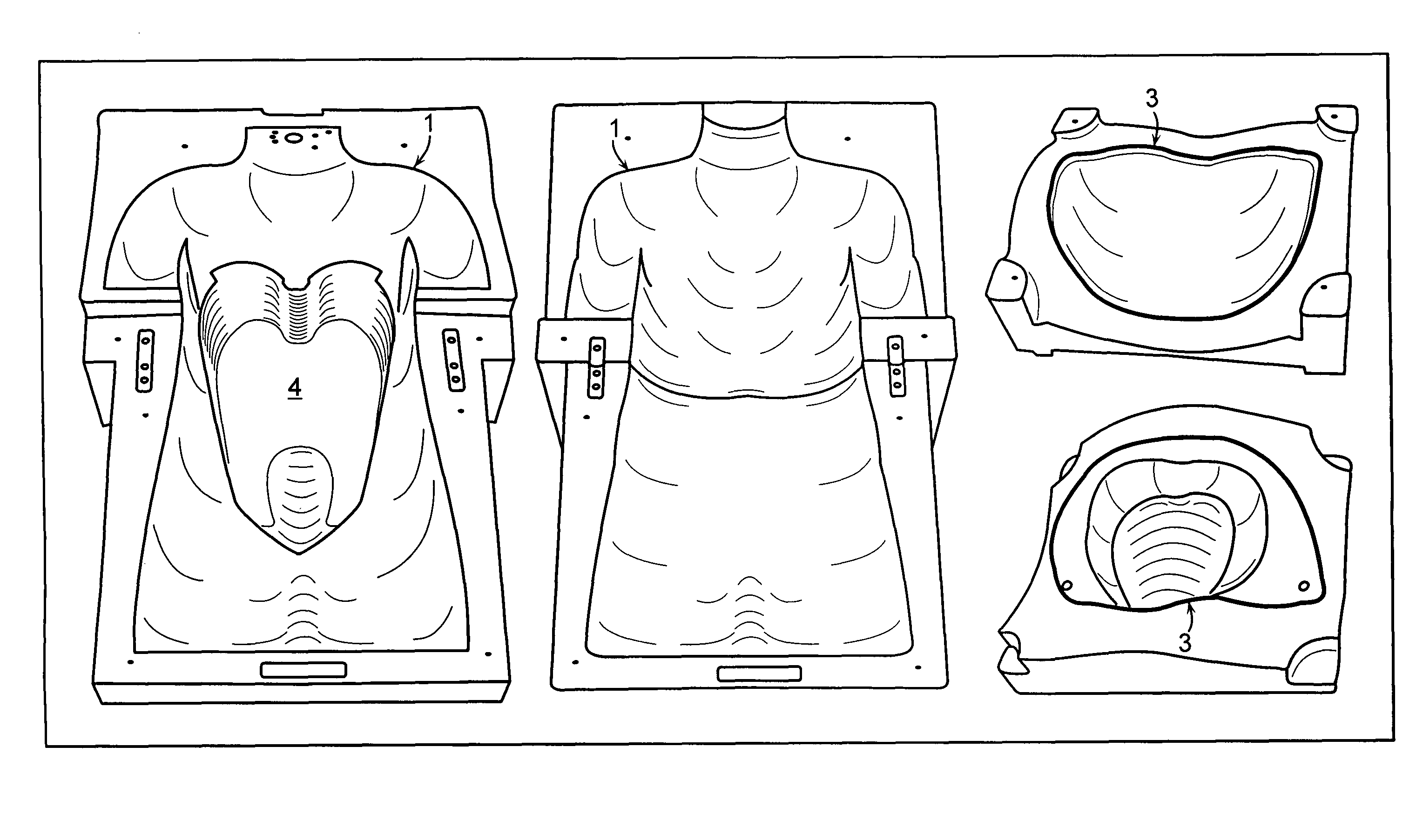
[0074]The synthetic torso formed in accordance with FIGS. 2-6 and 8 was used and evaluated by medical students, residents, and attending urologists who compared it to the standard training box. The study showed that the synthetic torso gives a more realistic approximation of a real procedure and is particularly suited for laparoscopy training. Animal organs were used in the studies to allow for operating on real tissues. The organs were placed in situs, requiring appropriate instrument access and port placement. Further, induced respiration caused the organs to move as in a live human body.

[0075]The trainee underwent numerous steps, beginning with the insertion of the Veress needle, insufflating the CO2, determining the port sites and placing the trocars. The trainee then performed a variety of laparoscopic procedures that necessitated dissecting and developing tissue planes, excising and reconstructing tissue, suturing, electrocautering, and performing other surgical maneuvers in a...


PUM


Abstract

A training and / or evaluating device is provided particularly useful in performing laparoscopic procedures, radiological procedures, and precise surgeries that simulates the structure and dynamic motion of the corresponding anatomical structure on which the procedure takes place. The device includes an outer housing, which may be designed to mimic the body wall, in which one or more organs are located. Motion of the organ(s), as a result of respiration, pulmonary action, circulation, digestion and other factors present in a live body, is simulated in the device so as to provide accurate dynamic motion of the organs during a procedure.

Description

[0001]The present application claims the benefit of U.S. provisional application No. 60 / 551,090 filed Mar. 8, 2004, which is incorporated herein by reference in its entirety.FIELD OF INVENTION[0002]In one aspect, the present invention relates to devices and methods for performing and evaluating surgical and other medical treatment procedures, including anatomical structures and methods for use in training and evaluating individuals in laparascopic surgeries. The devices and methods may further be used in training and evaluation of non-laparascopic surgeries, particularly those that require precision and are complicated by the dynamic motion of the organs and body parts acted upon during the procedure. The devices and methods may further be used in radioscopic training and evaluation. In preferred aspects, devices of the invention can mimic anatomical, dynamic and / or mechanical properties of that area of the human anatomy on which training and evaluation is being performed.BACKGROUND...

Claims


Application Information

Patent Timeline
Patent Type & Authority Patents(United States)
IPC IPC(8): G09B23/28
CPCG09B23/28
Inventor STOIANOVICI, DANMAZILU, DUMITRUPATRICIU, ALEXANDRUKAVOUSSI, LOUIS R.
Owner THE JOHN HOPKINS UNIV SCHOOL OF MEDICINE
Why Eureka
Eureka Blog
Learn More