
Computer-aided pathological diagnosis system

a computer-aided, pathological technology, applied in the field of medicine, can solve the problems of only local (single pixel) information, yield subjective and imprecise results, and current imaging practices are mostly manual, and achieve the effect of accurate identification of cell features

Active Publication Date: 2011-12-13
UNIV OF SOUTH FLORIDA
View PDF11 Cites 14 Cited by
  • Summary
  • Abstract
  • Description
  • Claims
  • Application Information

AI Technical Summary

Benefits of technology

The method provides accurate and objective classification of cancer cells, reducing false-positive and false-negative diagnoses, and improves cytology / pathology evaluations by enhancing cellular feature extraction and segmentation, thus aiding in better patient management.

Problems solved by technology

Current imaging practices are mostly manual, time-consuming, and tedious, yielding subjective and imprecise results.
The problem with these approaches is that they employ only local (single pixel) information.
Cellular images share the following characteristics:(1) Poor contrast: Object (cell) gray levels may be close to that of the background.(2) Many cluttered objects (cells) in a single scene: This high number of occluding objects makes image segmentation difficult.(3) Low quality: Traditional staining techniques introduce a lot of inhomogeneity into the images, where not all of the parts of the same tissue are equally stained.

Method used


Image

  • Computer-aided pathological diagnosis system
  • Computer-aided pathological diagnosis system
  • Computer-aided pathological diagnosis system

Examples


Embodiment Construction

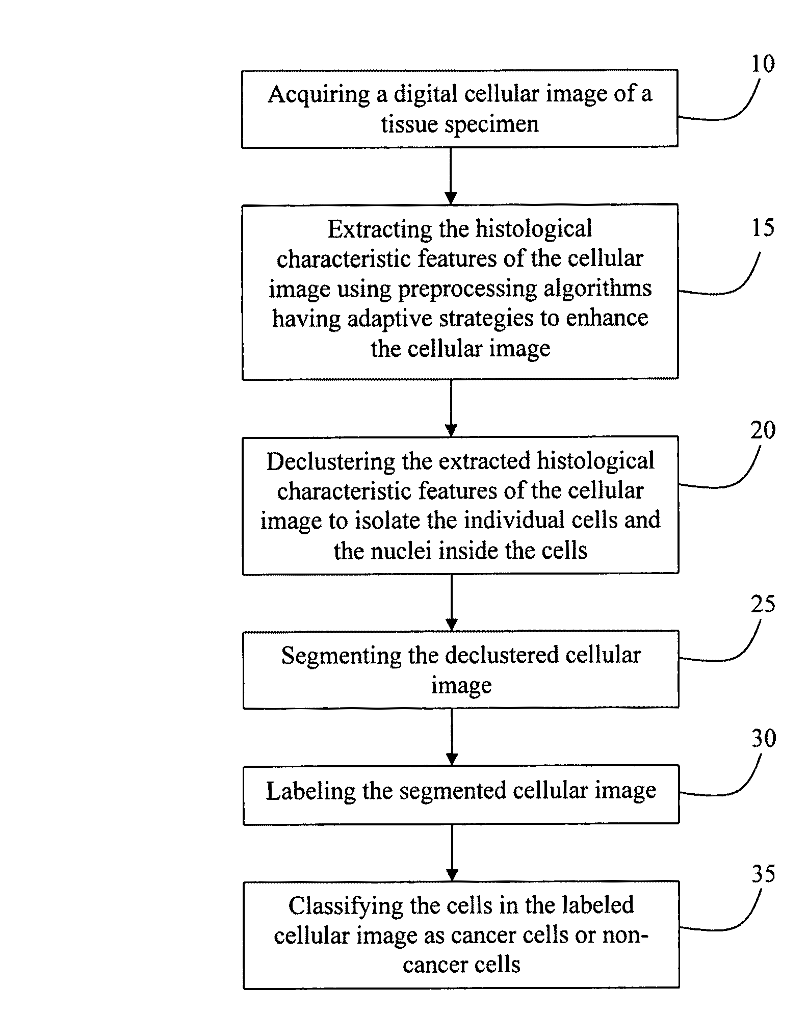
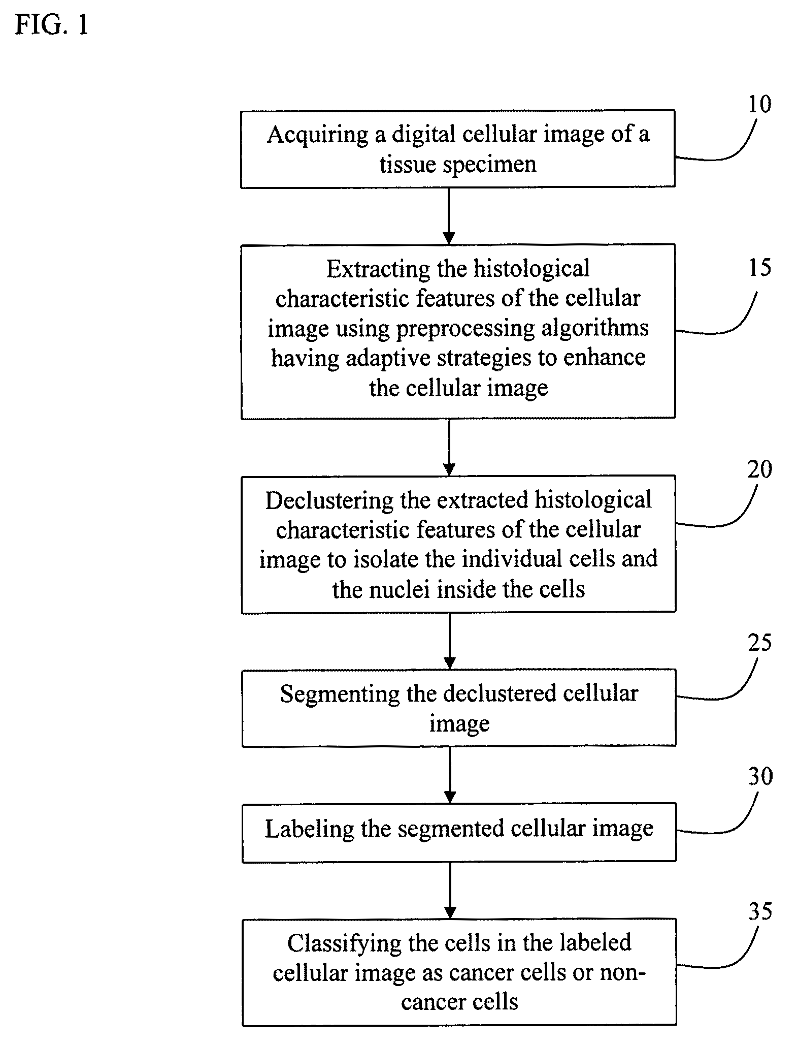
[0029]With reference to FIG. 1, the present invention is provides a computer-aided pathological diagnosis method for the classification of cancer cells in a tissue specimen based on a digital cellular image of the tissue specimen 10. The method of the present invention includes the steps of, extracting the histological characteristic features of the cellular image using preprocessing algorithms having adaptive strategies to enhance the cellular image 15, declustering the extracted histological characteristic features of the cellular image to isolate the individual cells and the nuclei inside the cells 20, segmenting the declustered cellular image 25, labeling the segmented cellular image 30 and classifying the cells in the labeled cellular image as cancer cells or non-cancer cells 35.

[0030]In a particular embodiment, the present invention is a Computer-Aided Pathological Diagnosis (CAPD) system designed for biomarker assessment, to differentiate lung cancer biomarkers and to identif...


PUM


Abstract

The present invention is a computer-aided pathological diagnosis method for the classification of cancer cells in a tissue specimen based on a digital cellular image of the tissue specimen. The method of the present invention includes the steps of, extracting the histological characteristic features of the cellular image using preprocessing algorithms having adaptive strategies to enhance the cellular image, declustering the extracted histological characteristic features of the cellular image to isolate the individual cells and the nuclei inside the cells, segmenting the declustered cellular image, labeling the segmented cellular image and classifying the cells in the labeled cellular image as cancer cells or non-cancer cells.

Description

CROSS-REFERENCE TO RELATED APPLICATIONS[0001]This application claims the benefit of U.S. provisional patent application No. 60 / 806,372, filed on Jun. 30, 2006, entitled, “Computer-Aided Pathological Diagnosis System”.STATEMENT OF GOVERNMENT INTEREST[0002]This invention was made with Government support under Grant No. PR054405 awarded by the Department of Defense and Grant No. SR33CA099046-01A1 awarded by the National Cancer Institute. The Government thus has certain rights in the invention.FIELD OF INVENTION[0003]This invention relates to the field of medicine. More specifically, this invention relates a computer-aided pathological diagnosis system designed for biomarker assessment, differentiation of cancer biomarkers and identification of cancer cells in tissue based upon an image of a specimen.BACKGROUND OF THE INVENTION[0004]Clinical lung cancer diagnosis depends upon the pathologist's interpretation of morphological features of histological and cytological specimens. These inte...

Claims


Application Information

Patent Timeline
Patent Type & Authority Patents(United States)
IPC IPC(8): G06K9/00
CPCG06T7/0012G06T2207/30024G06T2207/30061G06V20/695G06V20/698
Inventor QIAN, WEISONG, DANSHENGZHUKOV, TATYANA A.
Owner UNIV OF SOUTH FLORIDA
Learn More