
Ordered two-and three-dimensional structures of amphiphilic molecules

Inactive Publication Date: 2010-10-26
MOJTABAI FATEMEH
View PDF13 Cites 2 Cited by
  • Summary
  • Abstract
  • Description
  • Claims
  • Application Information

AI Technical Summary

Benefits of technology

[0009]The invention pertains, at least in part, to a new, simple and fast method for the fabrication of ordered structures, e.g., ordered two- and three-dimensional crystals, of amphiphilic molecules, such as but not limited to proteins, e.g., membrane proteins. Unlike conventional techniques, the methods of the invention allow for variation of the experimental parameters to facilitate fabrication of ordered structures with both long- as well as short-range orientation order, suitable for imaging techniques, such as high-resolution crystallographic analysis. In addition, the method is several times faster than conventional techniques, and maintains the native asymmetry of the amphiphilic molecule. The methods of the invention provide a general approach for crystallizing amphiphilic molecules such as membrane proteins. This is of considerable research interest since membrane proteins comprise a high proportion of pharmaceutically relevant targets. Yet the difficulties involved in crystallizing membrane proteins by using conventional techniques has posed a serious problem in the area of drug discovery. The present invention may lead to new discoveries of significant biotechnological importance.
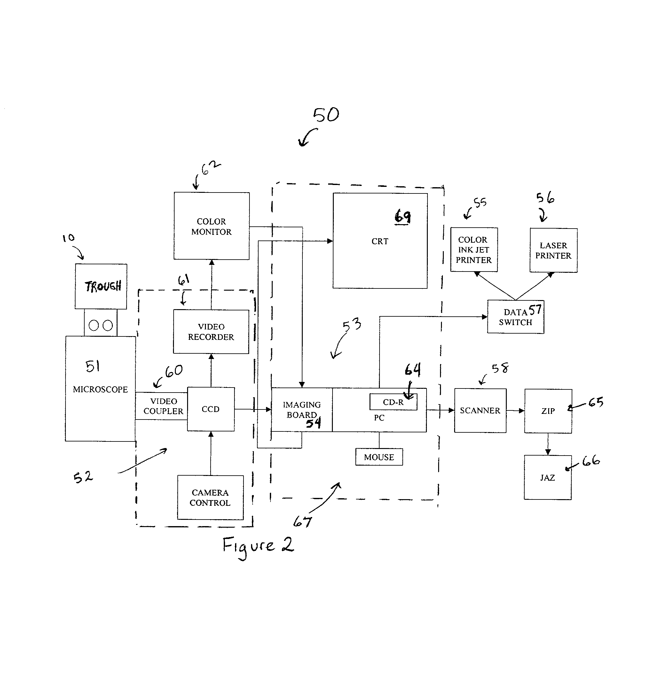
[0019]In one embodiment, the invention pertains to methods for forming ordered structures of amphiphilic molecules on an interface in a fast and controlled fashion. The methods may be performed using an apparatus with an integrated with real-time digital imaging laser fluorescence microscopy to allow for in-situ characterization of structure formation, throughout the fabrication process. The use of a robotics film transfer facilitates preparation of specimens for use in atomic resolution measurements such as electron-, x-ray crystallography as well as scanning probe microscopy.

Problems solved by technology

Yet the difficulties involved in crystallizing membrane proteins by using conventional techniques has posed a serious problem in the area of drug discovery.

Method used


Image

  • Ordered two-and three-dimensional structures of amphiphilic molecules
  • Ordered two-and three-dimensional structures of amphiphilic molecules
  • Ordered two-and three-dimensional structures of amphiphilic molecules

Examples


example 1

Direct Visual Evidence for the Formation of Ordered Structures, e.g., Large Ordered Domains, of a Membrane Proteins at an Air-Aqueous Interface

[0159]In this example, a unique ability to grow ordered structures of a membrane protein on a liquid in a fast and controlled fashion is demonstrated. The membrane protein cytochrome c oxidase (COX) from beef heart was chosen as a model membrane protein to elucidate the feasibility of the technique. The structure of COX has been extensively studied in different laboratories. The structure of the metal sites of COX was determined to 2.8 Å resolution by x-ray analysis (Tsukihara, T., et al. Science 269, 1069-74 (1995)). Crystal structure information at intermediate resolutions is available of tetragonal or hexagonal bipyramidal crystals that diffract x-rays to 5 Å (Shinzawa-Itoh, K., et al. J. Mol. Biol. 246, 572-575 (1995)) and 8 Å (Yoshikawa, S. et al. PNAS, 85, 1354-1358 (1988); Yoshikawa, S., et al. J. Crystal Growth, 122, 298-302 (1992)) r...

example 2

Electron Microscopy and Electron Diffraction of Ordered Domains Prepared at an Air-aqueous Interface

[0182]Ordered structures of the protein COX were formed at an air-aqueous interface for structure analysis by electron crystallography. The ordered structures were transferred to hydrophobic (carbon-coated) or hydrophilic (silicon oxide coated) grids. Two different types of specimen preparations were used for comparative analysis. In certain specimens, a complex was made between the protein, B-CC and FITC-SA. However, these probes were excluded in a majority of specimen preparations in order to eliminate the effect of bound ligands on the structure of the protein. In all specimen ordered structures, the lipids were probed with the fluorescently tagged lipid R-PE. Inclusion of the lipid probe facilitated visual monitoring of the ordered structure during its transfer to the grids. The electron microscope studies reported here were done on unstained specimen, at room temperature (22±2° C...

example 3

Direct Visual Evidence for Formation of Ordered Two-dimensional Ordered Structures of the Multidrug Resistance P-glycoprotein

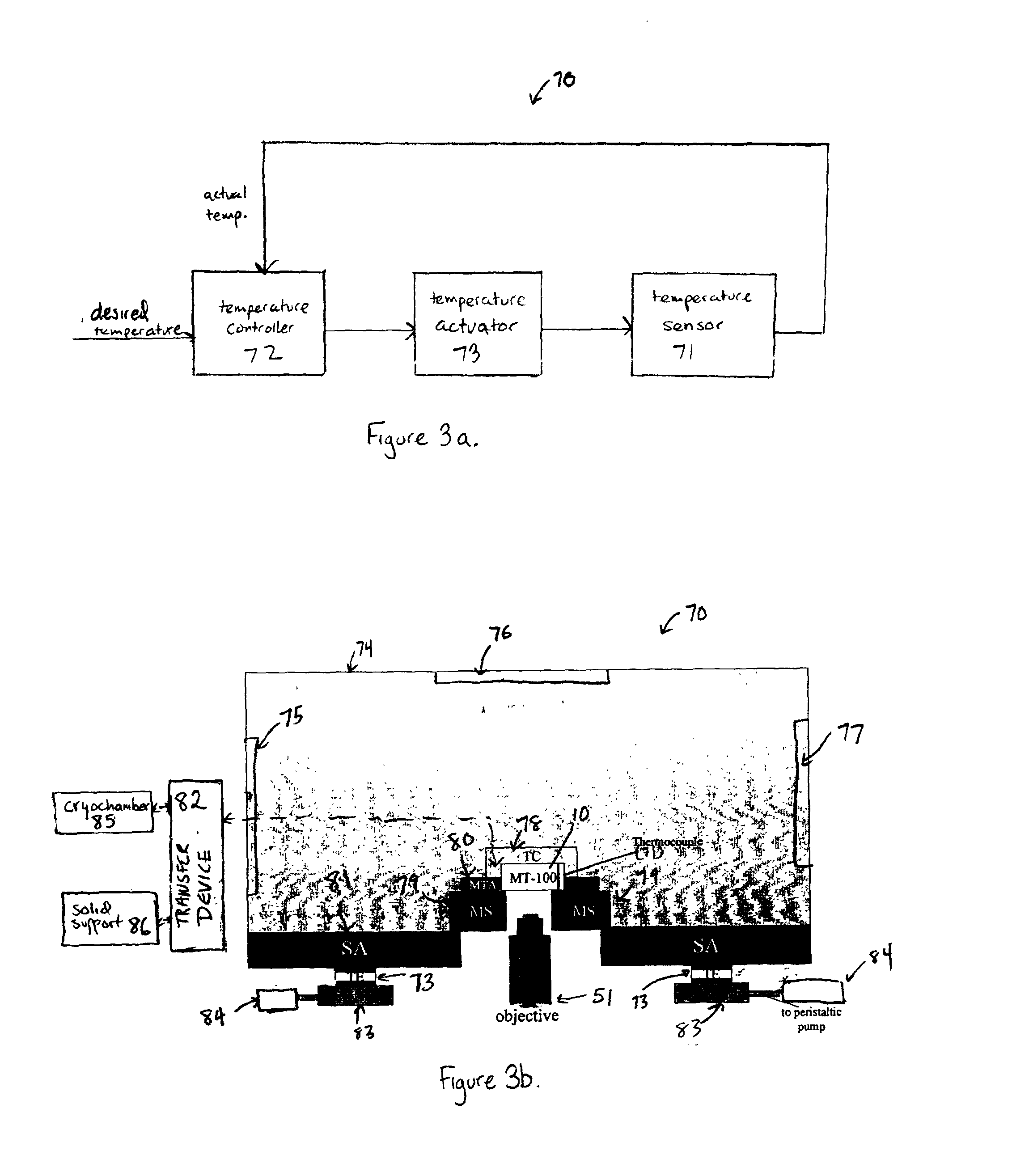
[0190]In another system, comprised of the multidrug resistance P-glycoprotein (P-gp), the formation of large ordered domains of the protein in its natural membrane was demonstrated. Non-random distribution of cell surface P-glycoprotein (P-gp) in monolayers spread from multidrug resistant MCF-7 AdrR and MCF-7 sensitive cell lines was shown. Cell membrane vesicles were prepared according to the method of Cornwell et al. (J. Biol. Chem. 261, 7921-28 (1986)). Monolayers were spread from vesicles in a hypertonic buffer including a high concentration of sucrose. The vesicles were spread at the air-aqueous buffer interface of the Micro-Trough MT-100, on a hypotonic buffer in the same manner as it was discussed earlier for COX. Spreading of the monolayer was monitored by observing an increase in the surface pressure of the spread film at a constant area. P-gp was pro...


PUM

PropertyMeasurementUnit
Areaaaaaaaaaaa
Kinematic viscosityaaaaaaaaaa
Surface energyaaaaaaaaaa

Abstract

The invention pertains, at least in part, to a method for forming an ordered structure of amphiphilic molecules, such as proteins. The method includes contacting a population of amphiphilic molecules with a interface; compressing said population laterally to an appropriate pressure, such that an ordered structure at the interface is formed. The invention also pertains to the two- and three-dimensional ordered structures that are formed using the planar membrane compression method of the invention.

Description

RELATED APPLICATIONS[0001]This application claims priority to U.S. Provisional Patent Application Ser. No. 60 / 242,913, entitled “Controlled Fabrication of Crystals and Long-Range Ordered Arrays of Membrane Proteins for High Throughput Structure Analysis and Screening Applications,” filed on Oct. 24, 2000; the entire contents of which are hereby incorporated herein by reference.BACKGROUND OF THE INVENTION[0002]Protein structure information is indispensable for the design of effective drugs. Designers need detailed structures of proteins, to atomic resolution, so that they can tailor their drugs to interact with specific target areas in a protein molecule. Conventionally, x-ray crystallography has been used to elucidate the three-dimensional (3-D) structure of proteins. This technique can accurately identify the location of atoms by diffracting x-rays from innumerable protein molecules stacked up in an ordered form, so called “crystal”. However, crystallographers have yet to overcome ...

Claims


Application Information

Patent Timeline
IPC IPC(8): C40B40/10C07B61/00C40B60/14
CPCB82Y15/00C07K1/113B01J2219/00317B01J2219/00585B01J2219/00596B01J2219/00659B01J2219/00725C07B2200/11C40B40/10C40B60/14G01N2500/20G01N33/6845
Inventor MOJTABAI, FATEMEH
Owner MOJTABAI FATEMEH
Learn More