
Optical method and system to determine distribution of lipid particles in a sample

a technology applied in the field of optical method and system for characterizing lipoproteins in samples, can solve problems such as particle separation

Inactive Publication Date: 2010-04-20
KIMBERLY-CLARK WORLDWIDE INC +1
View PDF13 Cites 8 Cited by
  • Summary
  • Abstract
  • Description
  • Claims
  • Application Information

AI Technical Summary

Benefits of technology

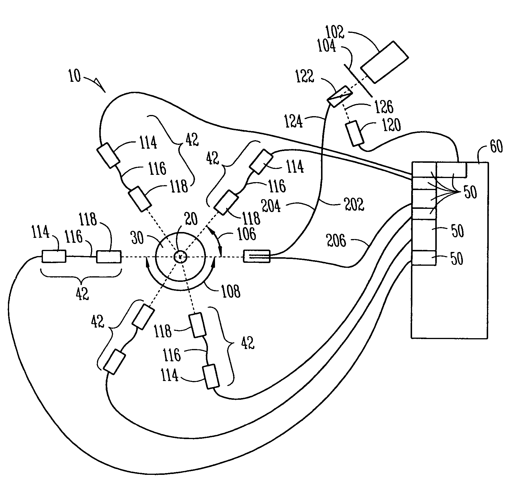
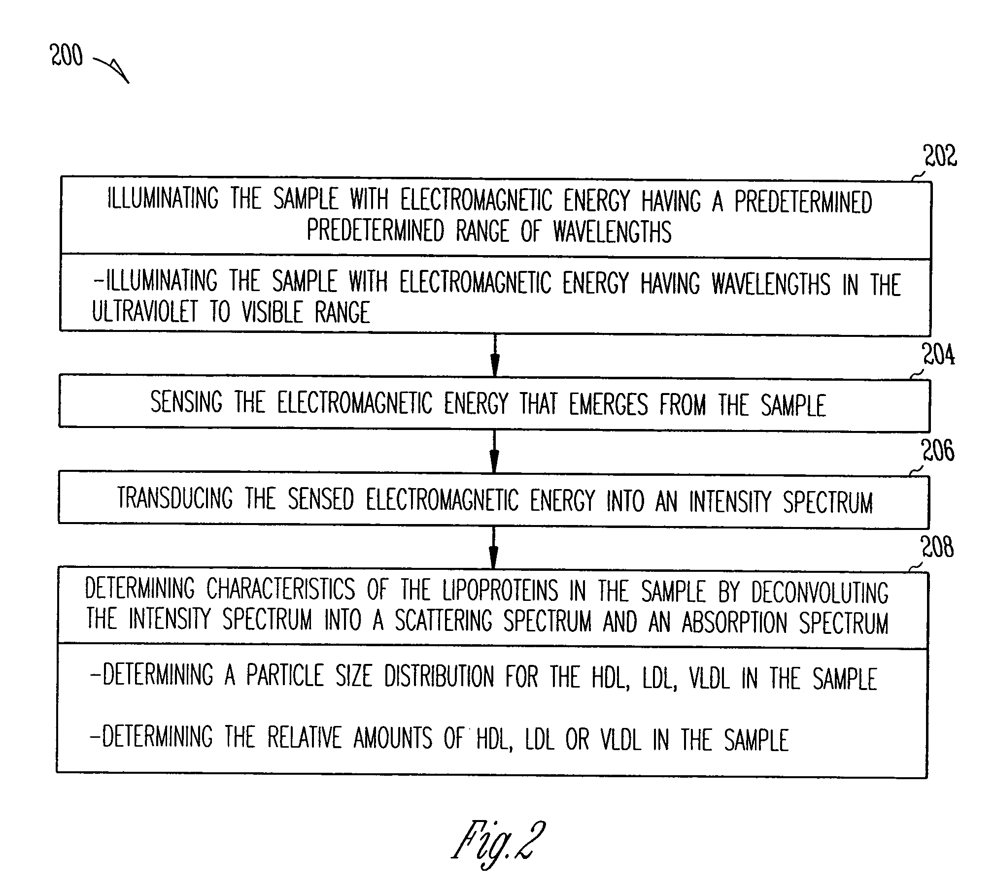
The present invention is a system and method for characterizing lipoproteins in a sample, such as blood. The system and method use absorption and scattering of electromagnetic energy by lipoprotein particles to provide a relative HDL / LDL / VLDL ratio and particle size distribution within each density class, as well as a semi-quantitative total cholesterol and triglyceride concentration. The system and method are effective because they may not require separation of the various classes of lipoprotein particles from whole blood or plasma. The analysis may be performed on small quantities of whole blood, plasma, or lipoproteins in saline. The system and method rely on the deconvolution of the angular and wavelength dependence of electromagnetic energy in the ultraviolet to visible range relative to scattering and absorption spectrophotometry. Different particle sizes show differences in the angular dependence of light scattering intensities, and differences in the type of lipoprotein may produce differences in the absorption spectrum. The system and method use a light source to illuminate the sample and sensors to sense the intensity spectrum which emerges from the sample. The intensity spectrum is then deconvolved to determine the types of lipoproteins in the sample. The method may include illuminating the sample at multiple observation angles and transducing the sensed electromagnetic energy into an intensity spectrum for each observation angle. The characteristics of the lipoproteins in the sample can be determined by deconvolving the intensity spectrum which emerges from the sample.

Problems solved by technology

However, these methods still require particle separation.

Method used


Image

  • Optical method and system to determine distribution of lipid particles in a sample
  • Optical method and system to determine distribution of lipid particles in a sample
  • Optical method and system to determine distribution of lipid particles in a sample

Examples


example

[0052]A purified fraction of Lp(a) particles was obtained in a 1 mL sample. Intensity spectra of the Lp(a) fraction were obtained by illuminating the sample as described above. The spectra were measured as a function of the particle concentration by recording in an Hp-8454 diode array spectrophotometer using a 1 cm path length micro-cuvette. Standard buffer saline was used as a diluent.

[0053]A set of optical properties was derived from the intensity spectra. The optical properties of the sample confirmed a portion of the theoretical optical properties that were previously determined.

[0054]FIG. 4 shows the measured spectra of the purified Lp(a) sample as function of concentration while FIG. 5 shows the normalized optical density spectrum for the sample. The normalization of the spectra eliminates the effect of the number of particles (as explained in C. E Alupoaei, J. A. Olivares, and L. H. Garcia-Rubio, “Quantitative Spectroscopy Analysis of prokaryotic Cells: Vegetative Cells and S...


PUM

PropertyMeasurementUnit
angleaaaaaaaaaa
sizeaaaaaaaaaa
path lengthaaaaaaaaaa

Abstract

The system and method of the present invention relate to characterizing lipoproteins in a sample. The system includes a light source that delivers electromagnetic energy in a predetermined range of wavelengths to the sample and a sensor that senses an intensity spectrum which emerges from the sample when the sample is illuminated by the light source. A processor determines a chemical composition of the sample to determine the presence of lipoprotein particles. The processor then characterizes lipoproteins that are within in the sample by deconvoluting the intensity spectrum into a scattering spectrum and absorption spectrum. The method includes illuminating the sample with electromagnetic energy having a predetermined range of wavelengths and sensing the electromagnetic energy that emerges from the sample. The method further includes transducing the sensed electromagnetic energy which emerges from the sample into an intensity spectrum that determines the types of lipoproteins that are within the sample.

Description

[0001]This application claims priority to U.S. Provisional Application No. 60 / 626,045 filed Nov. 8, 2004 and U.S. Provisional Application No. 60 / 563,614, filed Apr. 20, 2004 all of which are incorporated herein by reference.BACKGROUND[0002]The present invention relates to a method and system for characterizing lipoproteins in a sample, and more particularly, for characterizing lipoproteins in blood using light scattering and absorption properties of lipoproteins.[0003]Absorption spectroscopy and light scattering methods are typically used to determine the composition and / or molecular weight of macromolecules in solution. Until recently absorption and light scattering have been considered to be separate and independent measurements even though the same physical phenomena often occur in practice for both techniques.[0004]Absorption and light scattering measurements are usually conducted using electromagnetic energy having wavelengths in the ultraviolet to visible portion of the electr...

Claims


Application Information

Patent Timeline
Patent Type & Authority Patents(United States)
IPC IPC(8): G01N33/92G01N21/63G01N33/48G01N21/31G01N33/487
CPCG01N21/31
Inventor EVERHART, DENNIS S.LINDON, JACK N.GARCIA-RUBIO, LUIS
Owner KIMBERLY-CLARK WORLDWIDE INC
Learn More