
Methods for targeting and killing glioma cells

a glioma cell and glioma cell technology, applied in the field of anticancer pharmaceutical drugs, can solve the problems of limited application of oboc libraries, limited oboc diversity, and inability to routinely use analyses in clinical settings, so as to improve the effect of treatment effectiveness, reduce toxicities, and be well standardized and inexpensive

Inactive Publication Date: 2010-01-05
AUBURN UNIV
View PDF2 Cites 8 Cited by
  • Summary
  • Abstract
  • Description
  • Claims
  • Application Information

AI Technical Summary

Benefits of technology

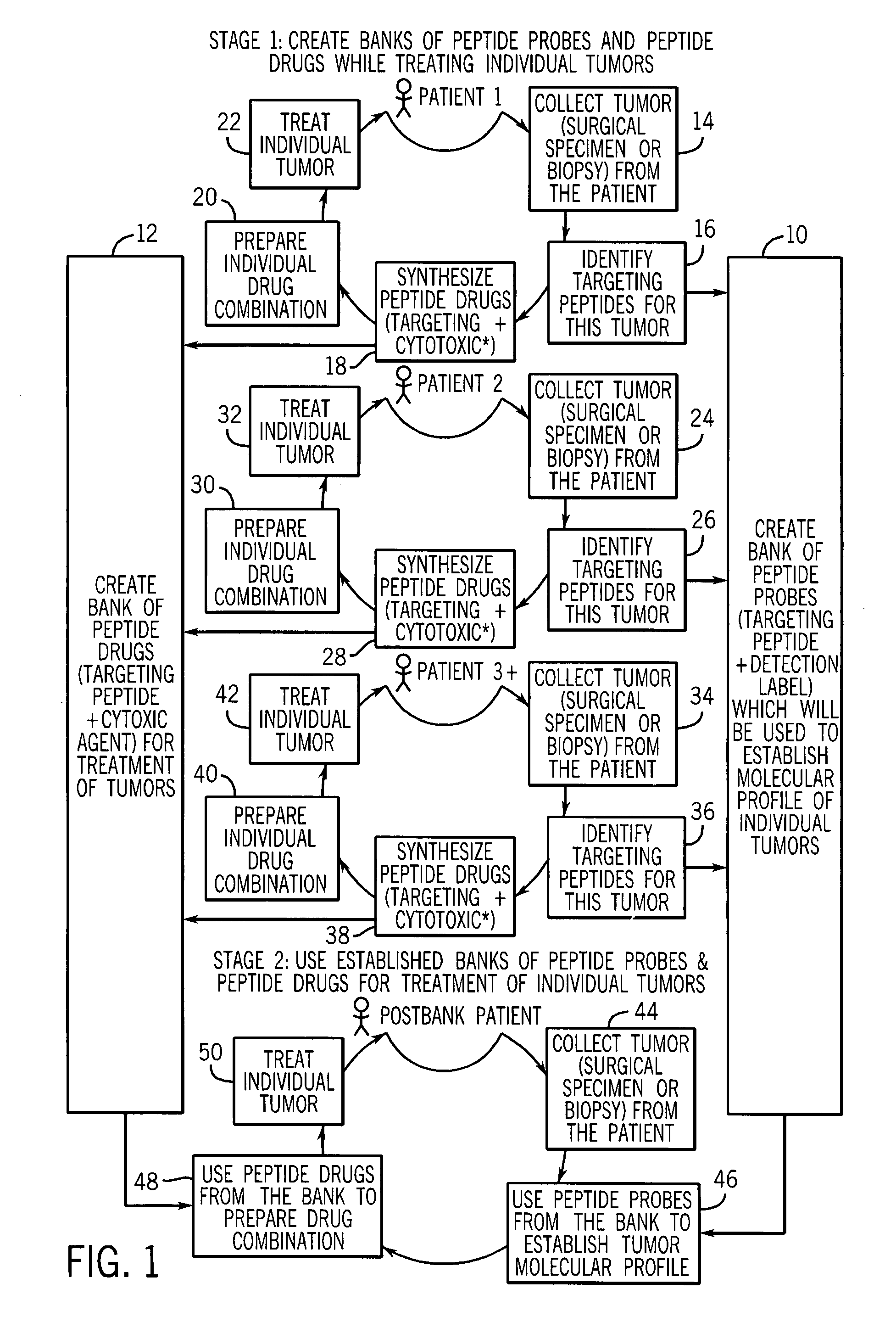
The patent describes a method for diagnosing and treating cancer by using targeting peptides that are specific to individual cancer cells. These peptides are obtained by screening phage display libraries with tumor cells and then using them to develop personalized anti-cancer drugs. The method can be used to design drugs that are effective against a wide range of cancers. The peptides are linked to a cytotoxic agent, which targets the tumor cells and induces a cytotoxic effect on them. The method can be used to treat cancerous tumors in humans and animals. The patent also describes a technique for identifying phage probes that target specific cancer cells and evaluating the heterogeneity of tumors from different patients. Overall, the method provides a more efficient and effective way to develop personalized drugs for cancer treatment.

Problems solved by technology

However, such analyses are unlikely to be used routinely in a clinical setting due to their complexity and the absence of 2-D protein databases of different tumor types.
While the method seems promising for profiling cancer cells derived from individual cancer specimens, the diversity of OBOC libraries is relatively low and could be a limiting factor for their broad applications.
In spite of the fact that some native cationic antimicrobial lytic peptides are more toxic to cancer cells than to normal mammalian cells, there are several limitations that prevent their application in cancer therapy.
The use of native all L-amino acid antimicrobial peptides in vivo is limited in part because of enzymatic degradation and binding to components in serum.

Method used


Image

  • Methods for targeting and killing glioma cells
  • Methods for targeting and killing glioma cells
  • Methods for targeting and killing glioma cells

Examples


examples

[0053]FIG. 4(a) shows three peptide families selected for rat brain tumor cells (glioma) (published in Samoylova et al, “Phage Probes for Malignant Glial Cells,” Molecular Cancer Therapeutics, pages 1129-1137 (2003)). The first family of peptides appeared to target a marker that is common for glioma cells, normal brain cells, and cells of non-brain origin. The second group or family of peptides contains peptides with pronounced glioma-selective properties (see FIG. 4(b)). Binding of phage expressing peptide with this consensus sequence to two gliomas was several magnitudes higher than binding to normal cells (see paper mentioned above). Because this peptide sequence was very selective for cancer cells, this peptide was used as the peptide later on as a targeting moiety to create an anti-cancer cytotoxic agent (see FIGS. 5(a) and (b)). The third family demonstrated 63-fold glioma selectivity when compared to normal brain cells, astrocytes.

[0054]Using glioma-selective sequences from t...


PUM

PropertyMeasurementUnit
concentrationaaaaaaaaaa
morphologyaaaaaaaaaa
sizeaaaaaaaaaa

Abstract

A diagnostic technique for matching anti-cancer peptide drugs to the molecular profiles of individual cancer tumors, and a method using the technique for a patient-specific treatment of tumors in mammals. This strategy matches anti-cancer peptide drugs to the molecular profiles of individual tumors, and includes the development of two banks, both of which use the same targeting peptides. Targeting peptides are identified using biopsies or other tissue materials from multiple cancer patients and placed into two banks. For a bank of probes, targeting peptides are linked to a label such as a fluorescent or a radioisotope. For a bank of drugs, targeting peptides are linked to a cytotoxic peptide to form a “targeting peptide drug”. Peptide probes are used to establish molecular profiles of individual tumors. Based on these molecular profiles, a patient-specific combination of targeting drugs from the bank of peptide drugs is prepared and the patient is treated. Any currently used cytotoxic agent that can be linked to targeting peptides may be used in place of cytotoxic peptides.

Description

CROSS REFERENCE TO RELATED APPLICATION[0001]The present application is based on and claims priority to U.S. Provisional Patent Application No. 60 / 572,511 filed May 19, 2004.FIELD OF THE INVENTION[0002]This invention relates to anti-cancer pharmaceutical drugs, and more particularly to a diagnostic technique for matching anti-cancer peptide drugs to the molecular profiles of individual cancer tumors, and to a method of using the technique for patient-specific treatment of such tumors in humans and animals.BACKGROUND OF THE INVENTION[0003]Cancer is a complex heterogeneous disease with molecular, morphological and clinical differences that exist both between and within tumors. While histopathologic features of tumor cell morphology, invasiveness and metastasis remain the “gold standard” for diagnosis and staging of cancers, molecular profiles of neoplastic cells based on DNA, mRNA, and / or protein alterations are rapidly being developed and utilized not only to augment diagnosis, but to...

Claims


Application Information

Patent Timeline
Patent Type & Authority Patents(United States)
IPC IPC(8): A61K38/17A61K51/00G01N33/574C10GG01N33/50
CPCG01N33/5011G01N33/574G01N2500/10
Inventor SAMOYLOVA, TATIANA I.BAKER, HENRY J.COX, NANCY R.GLOBA, LUDMILA P.MORRISON, NANCY E.
Owner AUBURN UNIV
Learn More