
Solid phase test device for sialidase assay

a sialidase and test device technology, applied in the field of enzyme assays, can solve the problems of increasing the risk of preterm birth and low weight infants, predicting adverse pregnancy outcome, and consuming time to complete the evaluation of bv by amsel criteria or by gram-stained smears, so as to facilitate analyte concentration and signal enhancement

Inactive Publication Date: 2009-09-22
ABBOTT RAPID DIAGNOSTICS INT UNLTD
View PDF23 Cites 7 Cited by
  • Summary
  • Abstract
  • Description
  • Claims
  • Application Information

AI Technical Summary

Benefits of technology

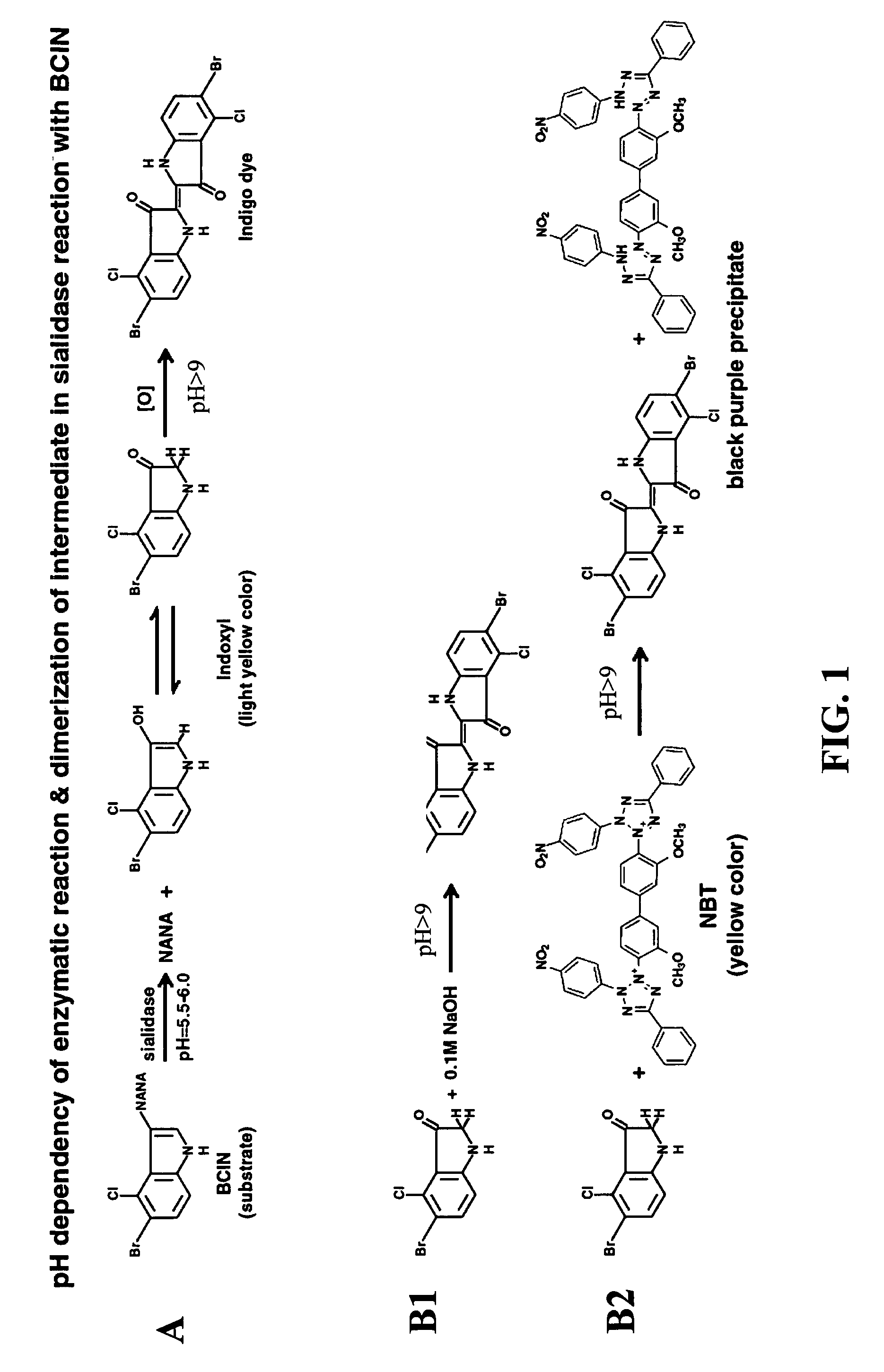
The present invention provides an improved method and devices for detecting sialidase activity in bodily fluids as a diagnosis tool for infectious diseases. The invention involves a solid-phase test device for detecting sialidase activity in a fluid sample, which comprises a sialidase detecting composition and a surfactant for intensifying the signal. The device can be used as a lateral flow sialidase assay or a spot test device. The method involves applying a fluid sample to the device and observing the appearance of a color change, which indicates the presence of sialidase in the sample. The invention can be used as a diagnostic tool for detecting infectious diseases associated with abnormal sialidase activity, such as Bacterial Vaginosis.

Problems solved by technology

Correlation was also found between elevated sialidase activities and an increased risk of preterm birth and low weight infants, suggesting that a sialidase activity test may predict adverse pregnancy outcome in women diagnosed with BV.
However, complete evaluation of BV by the Amsel criteria or by Gram-stained smears is time consuming.
Moreover, it requires skilled personnel and microscopic capabilities, which are not available in many of the physician offices or other clinic settings attended by women with BV.
As a result, samples need to be sent to a laboratory and results are further delayed, or more often, laboratory tests are not performed and diagnosis is based on clinical signs only, which may be misleading.

Method used


Image

  • Solid phase test device for sialidase assay
  • Solid phase test device for sialidase assay
  • Solid phase test device for sialidase assay

Examples


example 1

Preparation of Sialidase Test Strip

A. Preparation of NBT-Impregnated Sample Pads

[0042]Glass fiber filters (Millipore, GFCP0010000, 10 mm×10 cm) were soaked in NBT solution for 30 minutes in the dark at room temperature. The soaked glass fiber filters were placed on a blotting paper to remove excess fluids and then transferred to drying oven for 15 minutes at 50° C. The dried NBT-impregnated glass fiber filters (sample pads) were stored dried and dark in a dry room (RH 5-10%) at room temperature.

B. Card Assembly

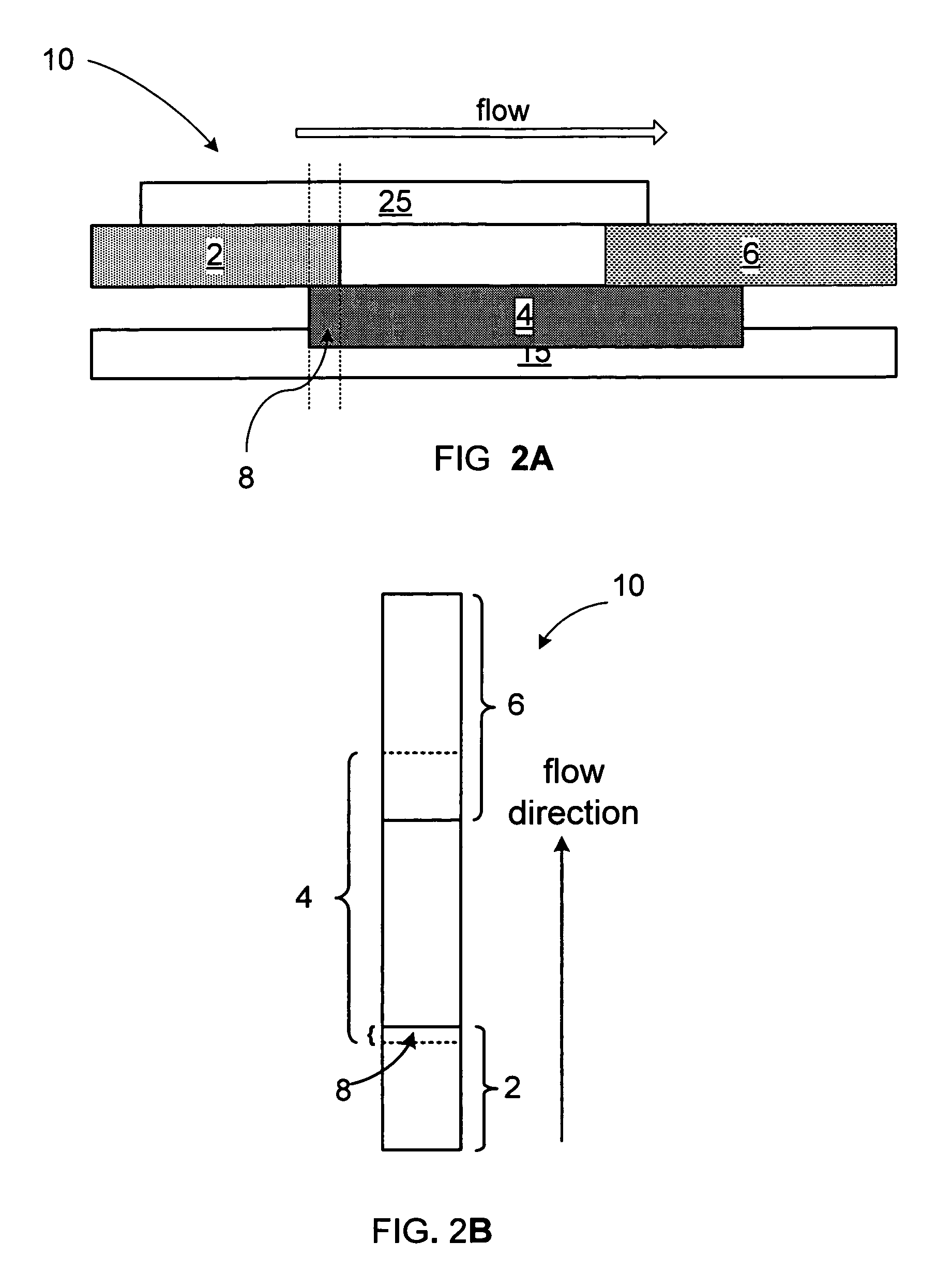
[0043]A test card was assembled according to the scheme in FIG. 2:[0044]1. A 43×250 mm piece of clear plastic film with a release liner protected adhesive, serving as the back laminate, designated 15 in FIG. 2, (ARcare 8876, Adhesive Research, Limerick, Ireland) was placed on top of a worktable. The release liner was peeled to expose the adhesive side of the tape.[0045]2. The reaction membrane (Nitrocellulose HF18004, Millipore, SA3J154101, 25×300 mm or Biodyne B, PALL, BNBZF3...

example 2

Running of Tests with Sialidase Test Strips

[0051]The strips constructed as described in example 1 above, were tested for sialidase activity with samples of sialidase producing bacteria: Bacteroides fragilis, sialidase negative bacteria: Lactobacillus plantarum and with purified sialidase.

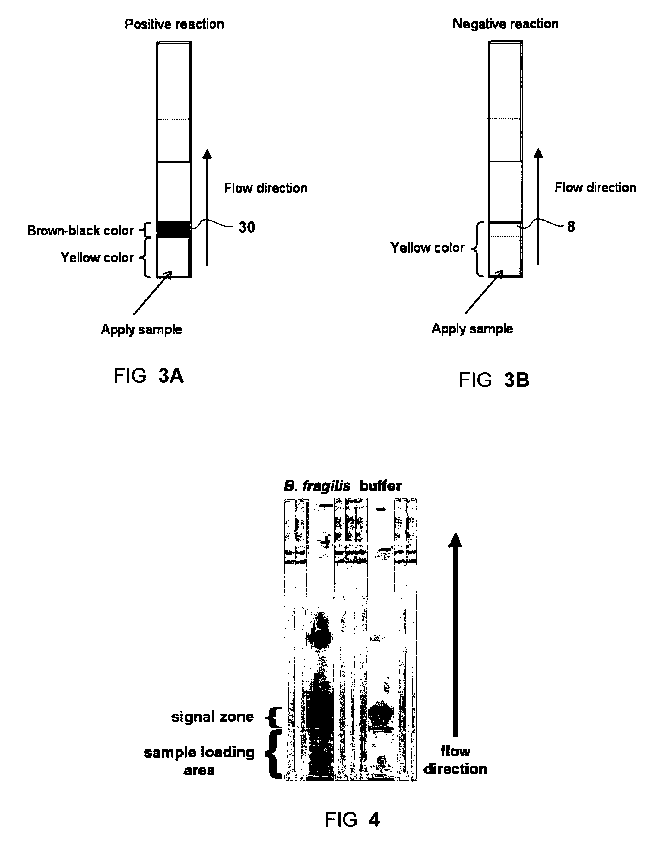
[0052]To start the test, 25 μl of sample was loaded onto the sample pad of the strip. The signal of positive sialidase reaction, a brown-purple color, was accumulated at the interface between the two different matrices (sample pad and the reaction membrane), namely the signal zone. Negative control (where no sialidase present) showed a yellow background at the signal zone. For each test the signal appearance time was recorded. The strips were observed up to 30 minutes. FIG. 4 shows exemplary results obtained with a sample of Bacteroides fragilis (left strip) and with running buffer (right strip) as a negative control.

[0053]The color change at the signal zone could be observed visually and was assign...

example 3

Clinical Samples (Vaginal Swabs)

[0055]47 clinical samples were tested to test the clinical relevance of the Sialidase test strip for the diagnosis of Bacterial Vaginosis BV. Vaginal discharge samples were obtained from volunteers at the Genitourinary Infections unit of the Wolfson Medical Center, Holon, Israel. Vaginal discharges were collected by a physician using a sterile swab (552C, Copan, Italia). The swab heads (tips) were placed in 2 ml screw-cap tubes and kept at 4° C. until use. The vaginal swabs were washed by adding 300 μl of running buffer in to the tube and by vortexing for 1 minute to elute the secretions from the swab and to achieve a homogenous sample. For each vaginal swab a diagnosis for BV was done using Gram staining and Nugent scoring. From the 300 μl swab wash, 25 μl were taken for the test. The test was done as described above for culture samples. Table 2 summarizes the result of 47 vaginal swabs washes that were diagnosed for BV and tested with the Sialidase ...


PUM

PropertyMeasurementUnit
lengthaaaaaaaaaa
pore sizeaaaaaaaaaa
temperatureaaaaaaaaaa

Abstract

A solid-phase test device and a method for the detection of sialidase activity in a fluid sample. The test device includes a solid support provided with a pre-deposited dry form of a sialidase detecting composition. The sialidase detecting composition includes a sialidase substrate and a color-developing reagent. When the sialidase substrate is exposed to sialidase it yields an intermediate compound which reacts with the color-developing reagent to form a detectable color change.

Description

BACKGROUND OF THE INVENTION[0001]1. Field of the Invention[0002]The present invention generally relates to enzyme assays and more specifically to a method and device for a dry format sialidase assay for the diagnosis of infectious diseases.[0003]2. Discussion of the Related Art[0004]The present invention discloses methods and devices for the detection of sialidase activity in fluid samples and in particular for the detection of sialidase activity in bodily fluids for the diagnosis of infectious diseases.[0005]Sialidases, also known as neuraminidases, are the enzymes that catalyze the cleavage of terminal sialic residues from carbohydrate moieties of glycoproteins, glycolipids, and proteoglycans. Sialidases specific for varying ketosidic linkages perform many biological functions. They are found in viruses, bacteria, parasites, and vertebrates, including mammals. Sialidases are associated with many diseases. Bacterial and viral sialidases may act as pathogenic factors in microbial in...

Claims


Application Information

Patent Timeline
Patent Type & Authority Patents(United States)
IPC IPC(8): G01N21/00
CPCC12Q1/34C12Q2334/50C12Q2334/52G01N2800/36G01N2333/30G01N2333/36G01N2333/195
Inventor DWIR, OREN SHRAGAKEREN, TOMERFISH, FALK
Owner ABBOTT RAPID DIAGNOSTICS INT UNLTD
Learn More