
Methods and compositions for detecting the activation state of multiple proteins in single cells

a technology of activation state and composition, applied in chemical methods analysis, instruments, biomass after-treatment, etc., can solve the problems of not being able to correlate rare subpopulations of cells, differences in cell physiology, and simultaneous detection of protein expression, protein form, and protein level for a multiplicity of proteins

Inactive Publication Date: 2009-07-21
THE BOARD OF TRUSTEES OF THE LELAND STANFORD JUNIOR UNIV
View PDF39 Cites 76 Cited by
  • Summary
  • Abstract
  • Description
  • Claims
  • Application Information

AI Technical Summary

Benefits of technology

"The present invention provides methods and compositions for simultaneously detecting the activation state of multiple activatable proteins in single cells using flow cytometry. This allows for the detection of protein activation patterns in cells for predicting or diagnosing disease states, and monitoring treatment progress. The methods involve contacting cells with a plurality of activation state-specific antibodies and substrates, and detecting the binding of the antibodies and the modification of the substrates in single cells. The invention also provides populations of cells, single cells, cell lysates, and proteins useful in the methods."

Problems solved by technology

To date it has not been possible to correlate rare subpopulations of cells with the activation state of kinases in important signaling pathways.
While random or misdirected proteolytic cleavage may be detrimental to the activity of a protein, many proteins are activated by the action of proteases that recognize and cleave specific peptide bonds.
This has largely been due to limitations imposed by the available investigative techniques.
It is well known that many proteins can function differently at different expression levels, and such differences in protein function can lead to differences in cell physiology.
However, even with the immobilization of a multiplicity of proteins from a sample, the problems of simultaneous detection of protein expression, protein form, and protein level for a multiplicity of proteins remains.

Method used


Image

  • Methods and compositions for detecting the activation state of multiple proteins in single cells
  • Methods and compositions for detecting the activation state of multiple proteins in single cells
  • Methods and compositions for detecting the activation state of multiple proteins in single cells

Examples


example 1

Functional Signaling Analysis in Single Cells: Simultaneous Measurement of Multiple Kinase Activities Using Polychromatic Flow Cytometry

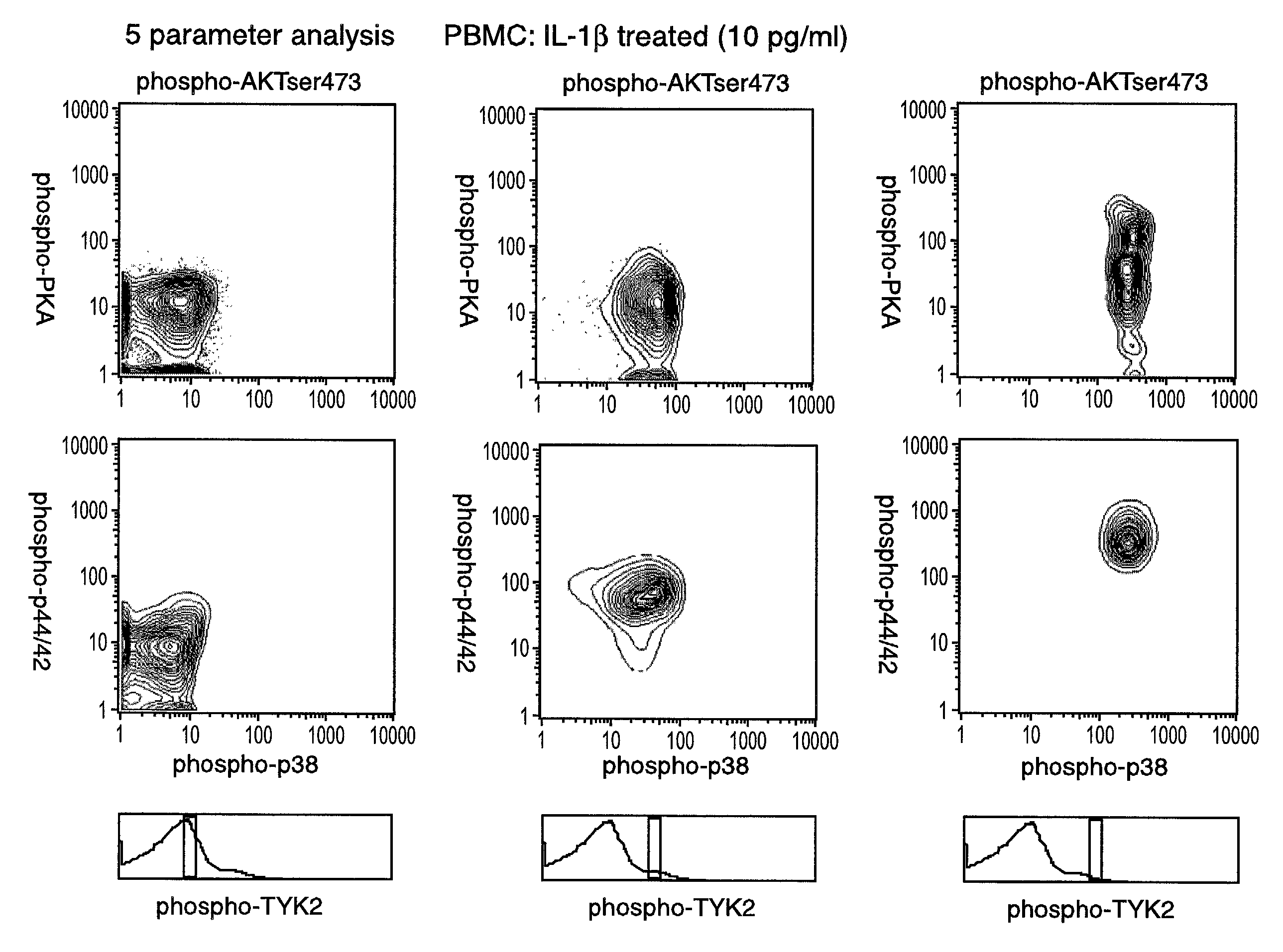
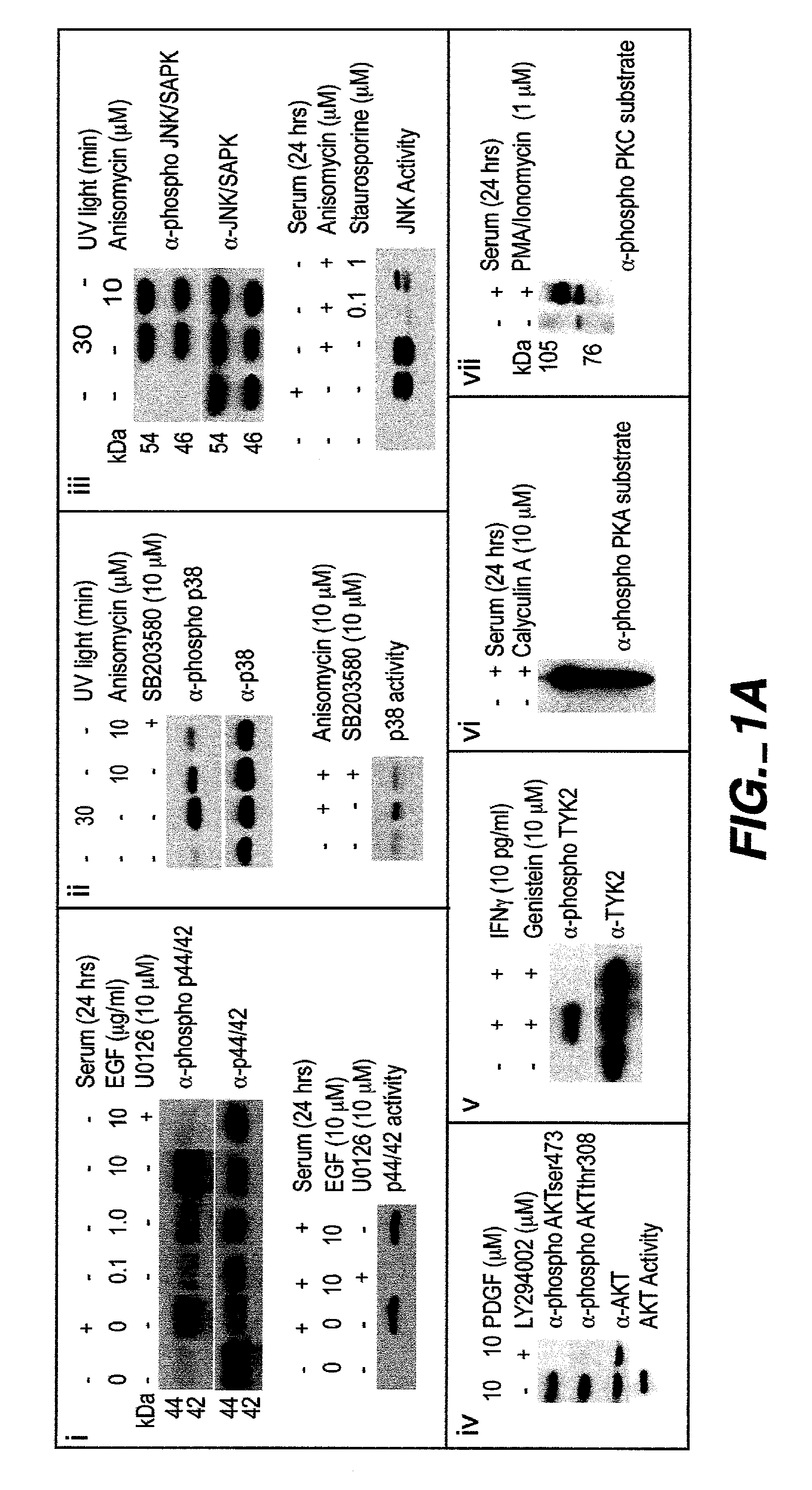
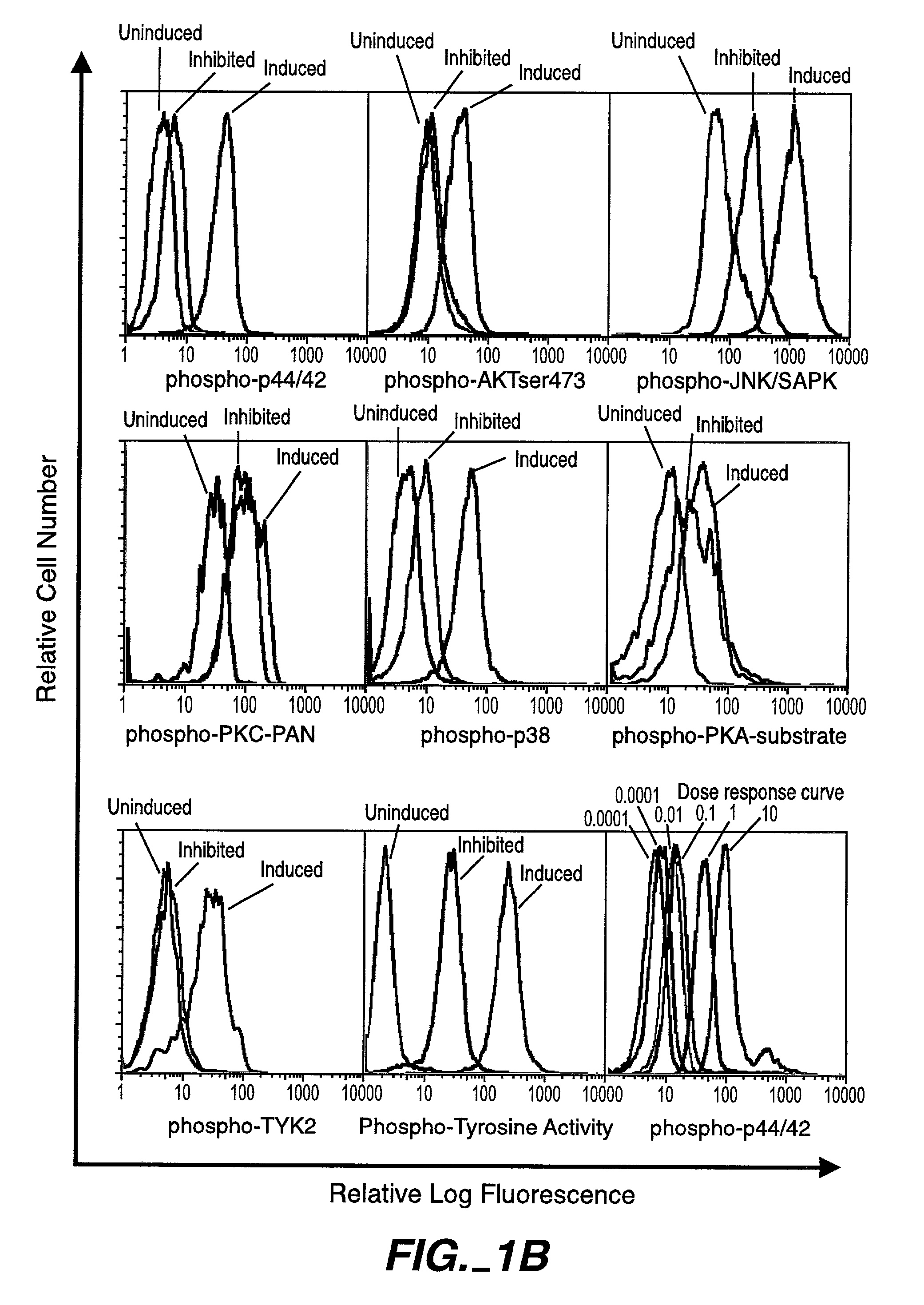
[0224]Intracellular assays of signaling systems has been limited by an inability to correlate functional subsets of cells in complex populations based on kinase activity. Such correlations could be important to distinguish changes in signaling status that arise in rare cell subsets during signaling or in disease manifestation. In this Example, using the methods and compositions of the present invention, the present inventors (also referred to herein as “we”) demonstrate the ability to simultaneously detect the kinase activities of members of the mitogen activated protein kineses family (p38 MAPK, p44 / 42 MAPK, JNK / SAPK), members of cell survival pathways (PKA, AKT / PKB), and members of T-cell activation pathways (TYK2, PKC) in subpopulations of complex cell populations by multi-parameter flow cytometric analysis. Further, in this Example the present i...

example 2

LFA-1 Functionally Contributes to T-Cell Activation and Polarized Effector Cell Cytokine Production to a TH1 Profile

[0283]In this Example, using the methods and compositions of the present invention, the present inventors (also referred to herein as “we”) demonstrate that LFA-1 stimulation by ICAM-2 functionally contributes in activating human naïve T-cells (CD3+CD4+CD8−CD62L+CD45RA+CD28+CD27+CD11adim) in the presence of CD3 / CD28 co-stimulation as determined by simultaneous multidimensional (33 parameters) assessment of immunophenotype, phospho-protein signaling, cytokine production, and transcriptional activity. Soluble ICAM-2 (sICAM-2), upon interacting with LFA-1, initiated phosphorylation and release of LFA-1 binding proteins cytohesin-1 and JAB1, which induced activation of the p44 / 42 MAPK pathway and cJun respectively. sICAM-2 enforced rapid kinetics of multiple active kinases (p44 / 42, p38, JNK MAPKs, Ick, AKT, PLCγ) when combined with CD3 / CD28 stimulation, lowering the CD3 / CD...

example 3

ICAM-2 / LFA-1 Activates p44 / 42 MAPK through PYK2 and SYK

[0360]In this Example, using the methods and compositions of the present invention, the present inventors (also referred to herein as “we”) demonstrate that Leukocyte Function Antigen-1-(LFA-1) was found to activate the RAS / RAF / MEK / MAPK cascade upon engagement with ICAM-2. Dissection of the signaling pathway in revealed the ICAM-2 / LFA-1 interaction depended on both PYK2 and SYK tyrosine kineses. Both PYK2 and SYK were found to associate with LFA-1 only after interaction with ICAM-2 and were rapidly re-distributed to the plasma membrane with concomitant phosphorylation of PYK2 and SYK as revealed by biochemical analysis and confocal microscopy. Chemical genetic approaches identify an LFA-1 mediated signal to phosphorylate PYK2, with subsequent signal relay to SYK to be contingent on PKC and PLCγ activities. Furthermore, cell-to-cell contact of ICAM-2+ cells with LFA-1+ initiated the transactivation of the p44 / 42 MAPK pathway in L...


PUM

PropertyMeasurementUnit
fluorescentaaaaaaaaaa
fluorescent activated cell sortingaaaaaaaaaa
conformational changeaaaaaaaaaa

Abstract

The invention provides methods and compositions for simultaneously detecting the activation state of a plurality of proteins in single cells using flow cytometry. The invention further provides methods and compositions of screening for bioactive agents capable of coordinately modulating the activity of a plurality of proteins in single cells. The methods and compositions can be used to determine the protein activation profile of a cell for predicting or diagnosing a disease state, and for monitoring treatment of a disease state.

Description

[0001]This application claims the benefit of U.S. provisional applications Ser. No. 60 / 304,434, filed Jul. 10, 2001, and Ser. No. 60 / 310,141, filled Aug. 2, 2001.[0002]The invention was made with United States Government support under Grant Numbers P01-AI39646, AR44565, AI35304, N01-AR-6-2227, and A1 / GF41520-01 awarded by the National Institutes of Health. The United States Government has certain rights in the invention.FIELD OF THE INVENTION[0003]The invention relates generally to the field of protein detection using flow cytometry. More specifically, the invention relates to simultaneously detecting the activation states of multiple proteins in single cells using flow cytometry and, more particularly, using polychromatic flow cytometry.BACKGROUND OF THE INVENTION[0004]Proteins are the major components of cells. The spatiotemporal expression pattern and the subcellular localization of proteins determines the shape, structure, and function of cells. Proteins are assembled from 20 di...

Claims


Application Information

Patent Timeline
Patent Type & Authority Patents(United States)
IPC IPC(8): G01N33/53C12Q1/00C12Q1/02C12Q1/37C12Q1/48G01N21/78G01N33/50G01N33/543G01N33/569G01N33/573
CPCC12Q1/485G01N33/5008G01N33/5041G01N33/5091G01N33/5094G01N33/54313G01N33/5302G01N33/582G01N33/573Y10T436/101666G01N2333/91205G01N2333/96466Y10S435/973Y10T436/25G01N21/6428G01N33/6845G01N2021/6441G01N2405/00G01N2440/14
Inventor PEREZ, OMAR D.NOLAN, GARRY P.
Owner THE BOARD OF TRUSTEES OF THE LELAND STANFORD JUNIOR UNIV
Learn More