Methods and reagents for the rapid and efficient isolation of circulating cancer cells

a technology reagents, which is applied in the field of methods and reagents for the rapid and efficient isolation of circulating cancer cells, can solve the problems of difficult detection and elimination of colonies, inability to treat all of them successfully, and efforts that have proved to be somewhat futil

Inactive Publication Date: 2008-02-19
MENARINI SILICON BIOSYSTEMS SPA
View PDF32 Cites 177 Cited by
  • Summary
  • Abstract
  • Description
  • Claims
  • Application Information

AI Technical Summary

Benefits of technology

[0030]The present invention provides a rapid and efficient screening method for the characterization of not only tumor cells, but also rare cells, or other biological entities from biological samples. The method of the invention provides highly sensitive analytical techniques which enable efficient enrichment for entities of interest. This two stage methodology which ensures enrichment of target bioentities while eliminating a substantial amount of debris and other interfering substances prior to analysis, allows for examination of sample sizes which would otherwise be impractical. The method described herein combines elements of immunomagnetic enrichment with multiparameter flow cytometric, microscopic and immunocytochemical analysis in a unique way. Other means of enrichment such as density gradient centrifugation or panning or alteration of target cell density by appropriate labeling may also be utilized. According to a preferred embodiment, the method of the invention enables assaying whole blood for cancer staging, monitoring and screening. The sensitive nature of the assay facilitates the detection of residual disease, thus making it possible to monitor for cancer recurrence.
[0033]In a further embodiment of the invention, the isolated cells are subjected to immunocytochemical analysis by flowcytometry or other analytical platforms. Such analysis facilitates diagnosis and provides important information to the clinician.

Problems solved by technology

Unfortunately, the metastatic colonies are harder to detect and eliminate and it is often impossible to treat all of them successfully.
These efforts have proven to be somewhat futile as the appearance of such antigens in blood have not been generally predictive and are often detected when there is little hope for the patient.
Despite the success of PSA testing, the test leaves much to be desired.
This is being done through the laborious procedure of isolating all of the mRNA from a blood sample and performing reverse transcriptase PCR.
158:326-337(1997)) no good correlation exists between the presence of such cells in blood and the ability to predict which patients are in need of vigorous treatment.
It is noteworthy that PCR is difficult, if not impossible in many situations, to perform quantitatively, i.e., determine number of tumor cells per unit volume of biological sample.
Additionally false positives are often observed using this technique.
There is an added drawback which is that there is a finite and practical limit to the sensitivity of this, technique based on the sample size examined.
As a further consideration, tumor cells are often genetically unstable.
Further, there are very considerable doubts in the medical literature regarding the existence of such cells and the potential of such information.
Also, there are doubts that the ability to detect cancer cells early on will give any useful information.
The extent of the magnetic field gradient within the test medium that may be obtained in such a system is limited by the strength of the magnets and the separation distance between the magnets.
One drawback of internal gradient systems is that the use of steel wool, gauze material, or steel microbeads, may entrap non-magnetic components of the test medium by capillary action in the vicinity of intersecting wires or within interstices between intersecting wires.
Various coating procedures have been applied to such internal gradient columns (see, e.g., U.S. Pat. No. 5,693,539 to Miltenyi and U.S. Pat. No. 4,375,407 to Kronick), however, the large surface area in such systems still creates recovery concerns due to adsorption.
Hence, internal gradient systems are not desirable, particularly when recovery of very low frequency captured entities is the goal of the separation.
Furthermore, they make automation difficult and costly.
Less obvious is the fact that the smaller the population of a targeted cell, the more difficult it will be to magnetically label and to recover.
Since mixing of cells with such particles for substantial periods of time would be deleterious, it becomes necessary to increase particle concentration as much a possible.
There is, however, a limit to the quantity of magnetic particle that can be added, as one can substitute a rare cell mixed in with other blood cells for a rare cell mixed in with large quantities of magnetic particles upon separation.
The latter condition does not markedly improve the ability to enumerate the cells of interest or to examine them.
There is another drawback to the use of large particles to isolate cells in rare frequencies (1 to 50 cells per ml of blood).
Despite the fact that large magnetic particles allow the use of external gradients of very simple design and relatively low magnetic gradient, large particles tend to cluster around cells in a cage-like fashion making the cells difficult to see or to analyze.
Hence, the magnetic particles must be released from the target cells before analysis, and releasing the particles clearly introduces other complications.

Method used


Image

  • Methods and reagents for the rapid and efficient isolation of circulating cancer cells
  • Methods and reagents for the rapid and efficient isolation of circulating cancer cells
  • Methods and reagents for the rapid and efficient isolation of circulating cancer cells

Examples


example 1

Formulation of Improved Magnetic Nanoparticles for the Efficient Isolation of Rare Cells from Whole Blood

[0071]Rare cells (e.g., tumor cells in patients with epithelial derived tumors, fetal cells in maternal blood or the like) can be present in frequencies below one rare cell per ml of blood. The number of blood smears required to detect such rare cells is prohibitively large. Assuming 10 rare cells in 10 ml of blood, which corresponds to 10 tumor cells in 5-10×107 white blood cells (leukocytes), cells can be transferred to a microscope slide by cytocentrifugation or by settling, stained with an antibody specific for the rare cells of interest and read manually or automatically. The maximum number of cells that can be transferred to one slide is about 500,000 cells which means 100-200 slides are required to process 10 ml of blood. The time required for analysis by this approach makes it impractical and economically unfeasible. Consequently, enrichment methods such as sample volume ...

example 2

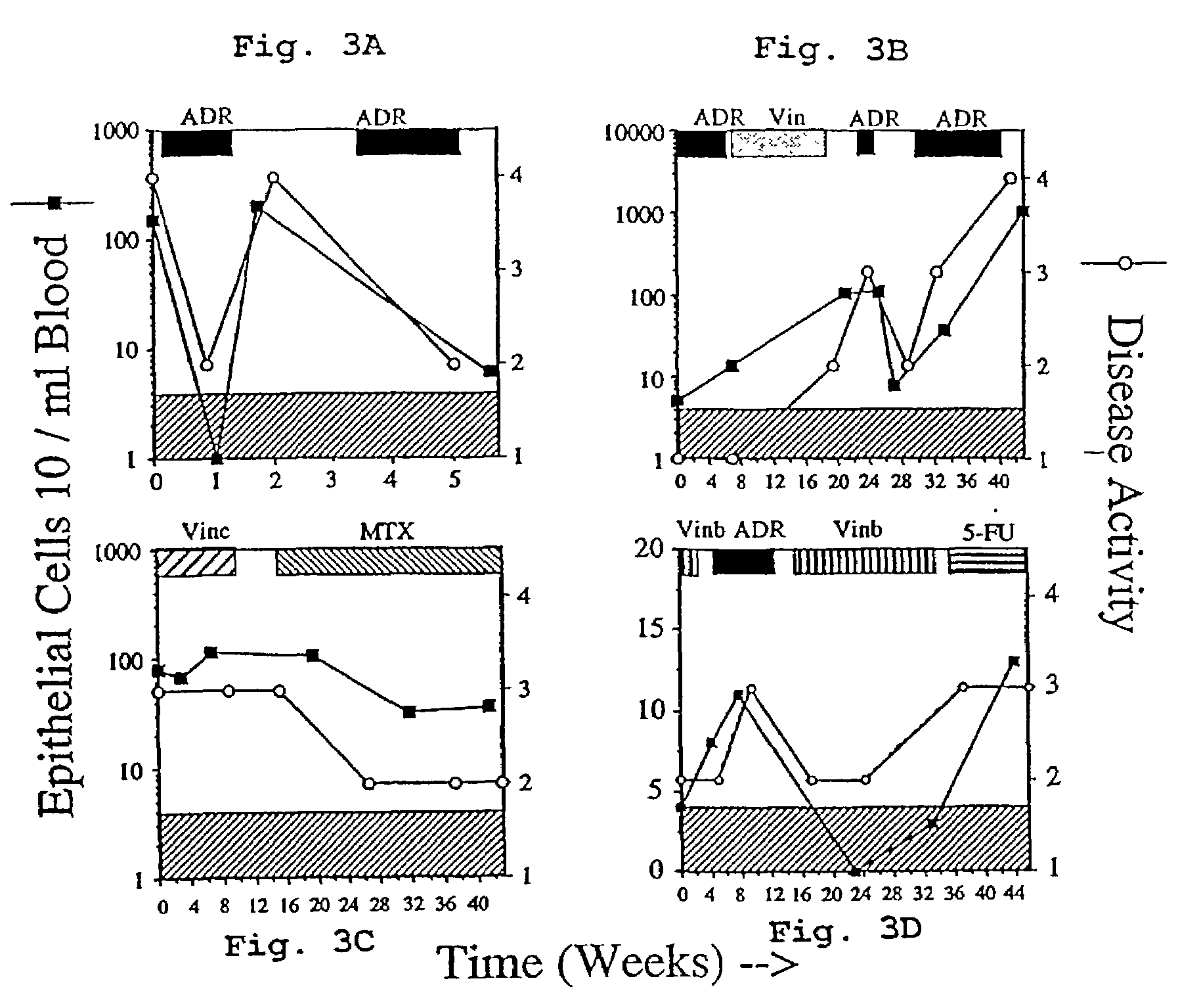
Enumeration of Circulating Epithelial Cells in Patients Treated for Metastatic Breast Cancer

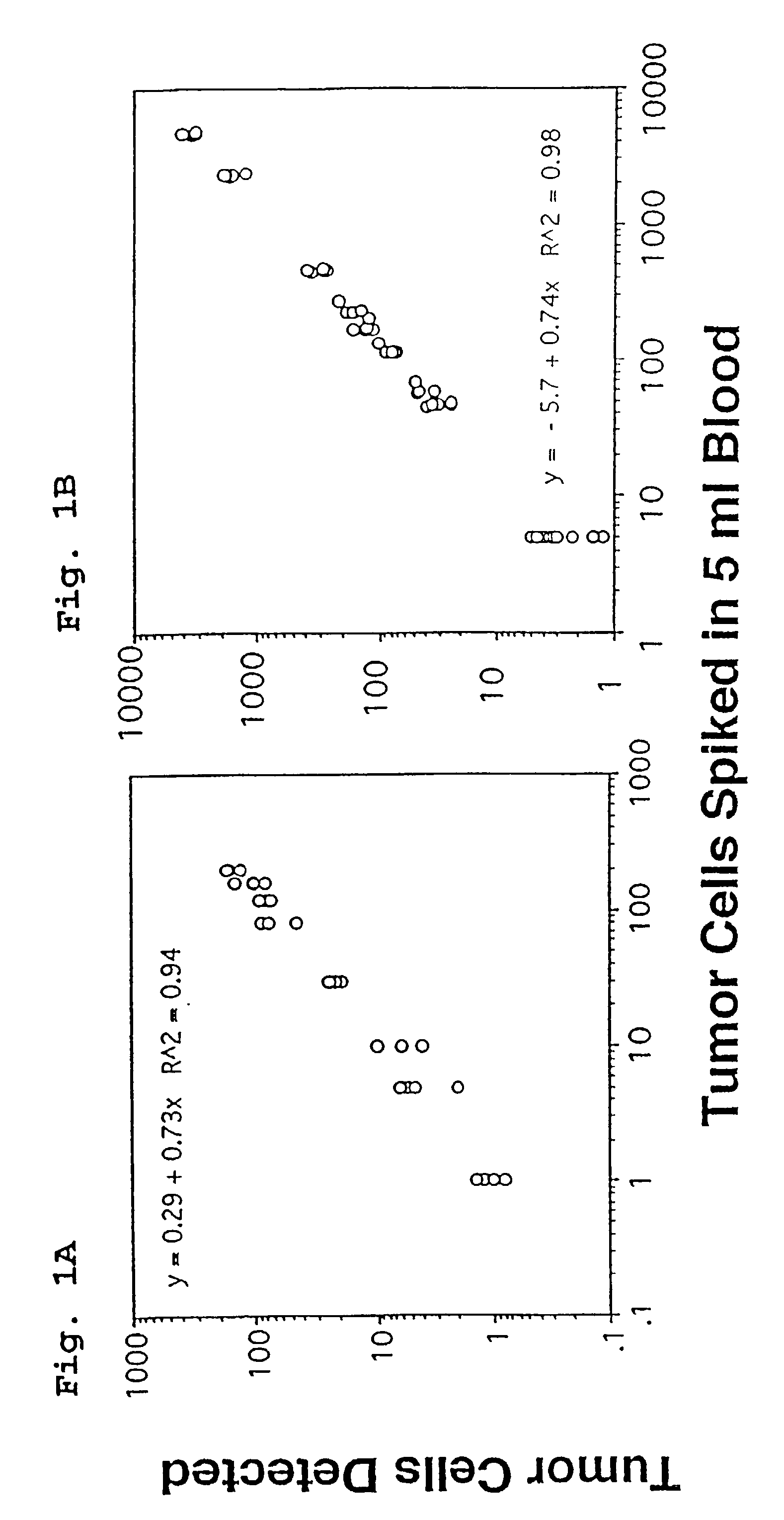
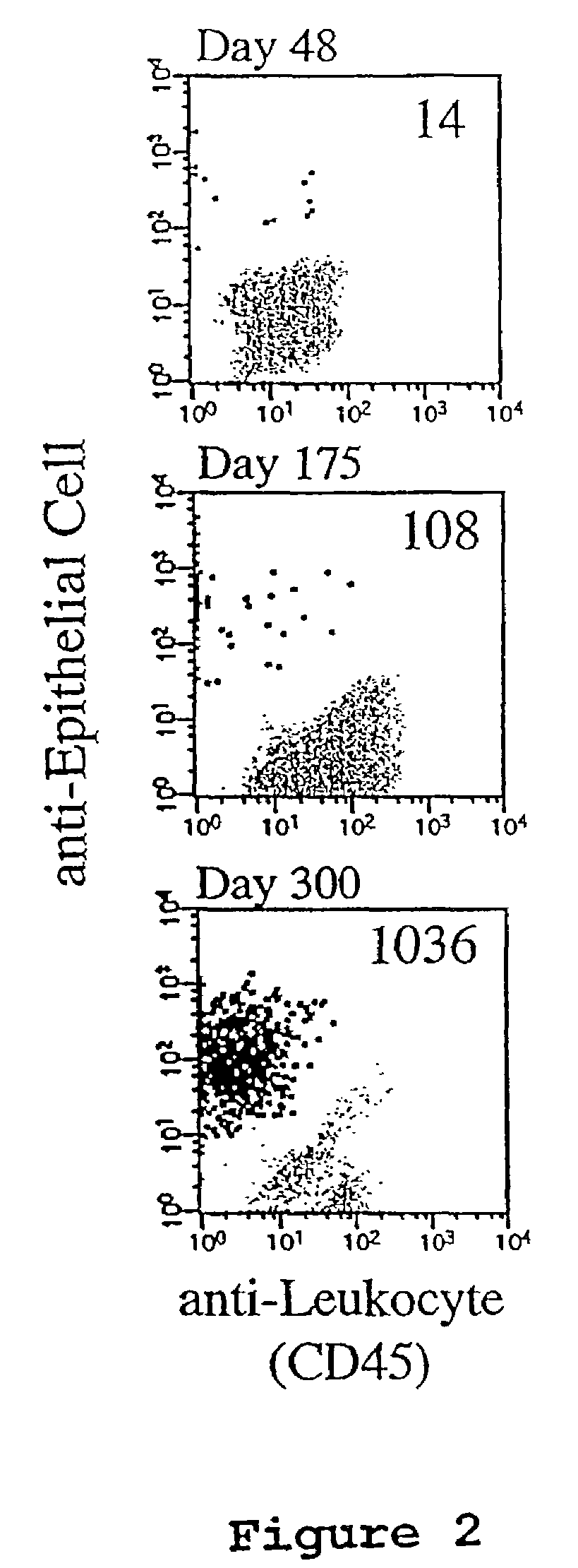
[0081]FIG. 1 shows the results obtained when tumor cells spiked into whole blood are isolated using the assay methods of the present invention. Panel A shows analysis by microscopy and panel B shows analysis results obtained using flow cytometry. FIG. 2 shows three examples of the flowcytometric analysis of 10 ml blood samples obtained from one patient with metastatic breast carcinoma at three time points, and includes the correlative display of the anti-leukocyte versus anti-epithelial cell antibodies of the flowcytometric analysis. In FIG. 2, Panel A, 14 events are detected and are present in the location typical for epithelial cells. In Panel B, 108 epithelial cells are detected and in Panel C 1036 epithelial cells are detected.

[0082]The number of events passing the threshold set on the nucleic acid dye in the analysis of the 10-ml blood sample varied between 5,000 and 50,000 events. These...

example 3

Enumeration of Circulating Epithelial Cells in Patients with No Evidence of Disease After Surgery for Carcinoma of the Breast with Curative Intent

[0100]Peripheral blood of 37 patients between 1 and 20 years after surgery was examined for the presence of epithelial cells by flowcytometry. Up to 7 peripheral blood samples were taken over a one-year period from these patients. In Table III, each of the patients is listed and sorted according to the TNM (tumor, node, and metastasis) stage at the time of surgery followed by the years after surgery. Table III also shows whether or not the patient received treatment (either chemotherapy or hormonal therapy) during the period studied. In 3 of 6 patients with evidence of distant metastasis in the past, but in complete remission at the time of study, epithelial cells were found in the blood at a higher frequency than that found in the control group. Circulating epithelial cells were also found in 9 of 31 patients with no evidence of distant m...


PUM

PropertyMeasurementUnit
Fractionaaaaaaaaaa
Fractionaaaaaaaaaa
Volumeaaaaaaaaaa

Abstract

A highly sensitive assay is disclosed which combines immunomagnetic enrichment with multiparameter flow cytometric and immunocytochemical analysis to detect, enumerate and characterize carcinoma cells in the blood. The assay can detect one epithelial cell or less in 1 ml of blood and has a greater sensitivity than conventional PCR or immunohistochemistry by 1-2 orders of magnitude. In addition, the assay facilitates the biological characterization and staging of carcinoma cells.

Description

CONTINUING APPLICATION DATA[0001]This application is a continuation of U.S. application Ser. No. 09 / 904,472, filed Jul. 13, 2001 now U.S. Pat. No. 6,645,731, which is a divisional application of U.S. application Ser. No. 09 / 248,388, filed Feb. 12, 1999, now U.S. Pat. No. 6,365,362, which in turn claims priority under 35 U.S.C. §119(e) to U.S. provisional application Nos. 60 / 074,535, filed Feb. 12, 1998, 60 / 110,279 and 60 / 110,202, each filed Nov. 30, 1998. Each of the above identified applications is incorporated by reference herein.FIELD OF THE INVENTION[0002]This invention relates to the fields of oncology and diagnostic testing. The invention is useful for cancer screening, staging, monitoring for chemotherapy treatment responses, cancer recurrence or the like. More specifically, the present invention provides reagents, methods and test kits which facilitate analysis and enumeration of tumor cells, or other rare cells isolated from biological samples.BACKGROUND OF THE INVENTION[00...

Claims


Application Information

Patent Timeline
IPC IPC(8): G01N33/53C07K1/13C07K17/00G01N33/546G01N33/553B03C1/01G01N33/58C12Q1/02C12Q1/06C12Q1/24G01N33/48G01N33/543G01N33/574H01F1/00
CPCB03C1/01B82Y25/00H01F1/0054G01N33/57484G01N33/57492G01N33/574G01N33/58G01N33/569G01N33/549
Inventor TERSTAPPEN, LEON W. M. M.RAO, GALLA CHANDRAUHR, JONATHAN W.RACILA, EMILIAN V.LIBERTI, PAUL A.
Owner MENARINI SILICON BIOSYSTEMS SPA
Why Eureka
Try Eureka