
Radioscopy using Kalpha gadolinium emission

a technology of kalpha gadolinium and kalpha, which is applied in the field of kalpha gadolinium emission radioscopy, can solve the problems of difficult to ensure high precision of radiation treatment, weakening of surrounding healthy tissues, and separation of stages in tim

Inactive Publication Date: 2008-01-01
KUMAKHOV MURADIN ABUBEKIROVICH
View PDF9 Cites 2 Cited by
  • Summary
  • Abstract
  • Description
  • Claims
  • Application Information

AI Technical Summary

Benefits of technology

"The patent proposes a method for radiation treatment of malignant neoplasms using X-ray beams for determining tissues structure and locating the malignant focus. The method involves introducing a gadolinium-containing preparation into the patient's body and detecting secondary radiation arising from the affected tissues. The detection is performed using a combination of X-ray beams and detectors sensitive to Kα-radiation of gadolinium atoms. The method allows for accurate determination of the malignant neoplasm's location and reduces the dose of radiation to healthy tissues. The technical effects of the patent include improved accuracy of malignant neoplasm location and reduced radiation dose to healthy tissues."

Problems solved by technology

Principal disadvantage of the above combination of patient preparation to irradiation and irradiation proper lies in the fact of the stages being separated both in time and in space, in particular, due to that they are accomplished using different means.
As a consequence, it is extremely difficult to ensure high precision of radiation treatment, with the result of malignant focus tissues remaining partially unirradiated or intensive X-ray radiation being concentrated in the region exceeding the malignant focus in size.
In the latter case, surrounding healthy tissues are damaged substantially stronger than at inevitable irradiation of healthy tissues located in the way of radiation to the malignant focus.
In realizing such technique, not only imprecision of the reference points selection and X-ray beams “aiming” during radiation treatment became manifest, but also variability of the internal organs location, and inaccuracy of patient accommodation during different seances of radiation treatment.
Moreover, dose fractionation in itself, though caused by endeavor to avoid overirradiation of healthy tissues, results in vicious circle, because it is known that dose required for irreversible damage of the malignant focus, when applied in single exposure, is several times as less as the total dose required at fractionation [1, p.84, 91].
However, none of such measures allow to avoid inaccuracies in the “aiming” of beams effecting radiation exposure on malignant neoplasm, caused by tumor growth.
At that, however, different beams are used for producing said images and for radiation treatment of malignant focus tissues, which allows not in principle to avoid mistakes in irradiating beams orientation.
In case of projections overlapping of the neoplasm and some solid organs, unequivocal conclusion is difficult to make.
Effect of drawbacks of the group considered is diminished in the means realizing computed tomography principles, which entails not only complexity of corresponding technical means, but also rather high irradiation dose.

Method used


Image

  • Radioscopy using Kalpha gadolinium emission
  • Radioscopy using Kalpha gadolinium emission
  • Radioscopy using Kalpha gadolinium emission

Examples


Embodiment Construction

[0060]The method proposed for determination of refined position of the malignant neoplasm is used as a stand-alone one if it is not followed with radiation treatment of the malignant neoplasm to accomplish damaging of its cells, or as a part of such treatment of the malignant neoplasm at the first stage of its realization. In both cases, this method as such is not a diagnostic or therapeutic one.

[0061]The method proposed of radiation treatment of the malignant neoplasm for damaging of its cells always comprises at the first stage of its realization a method proposed for position refinement of the malignant neoplasm.

[0062]Both methods abovementioned comprise method of gadolinium presence detection in tissues and organs of human body.

[0063]The device proposed is a common one for all methods.

[0064]The methods proposed are accomplished using the device proposed in a following way.

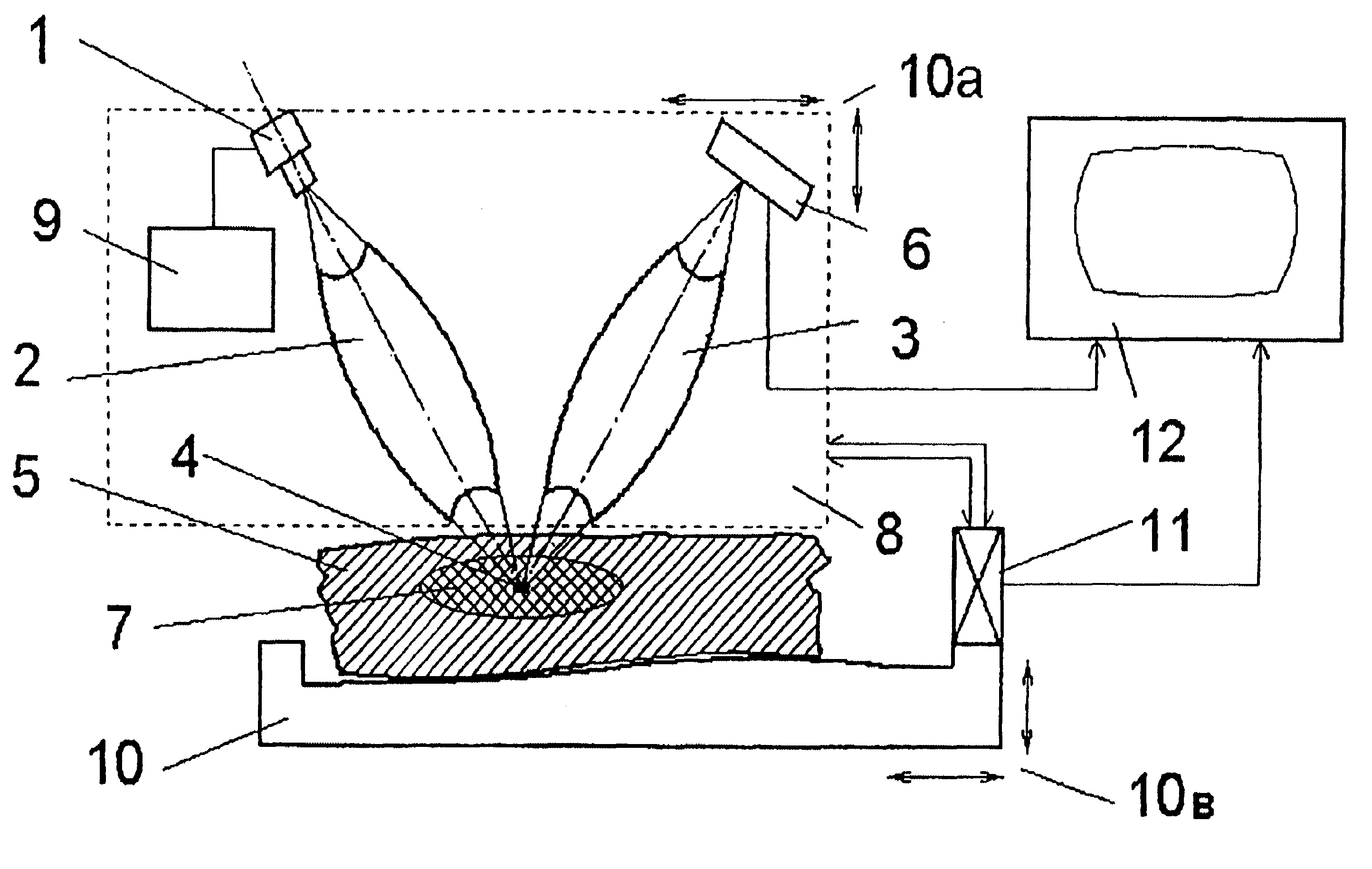
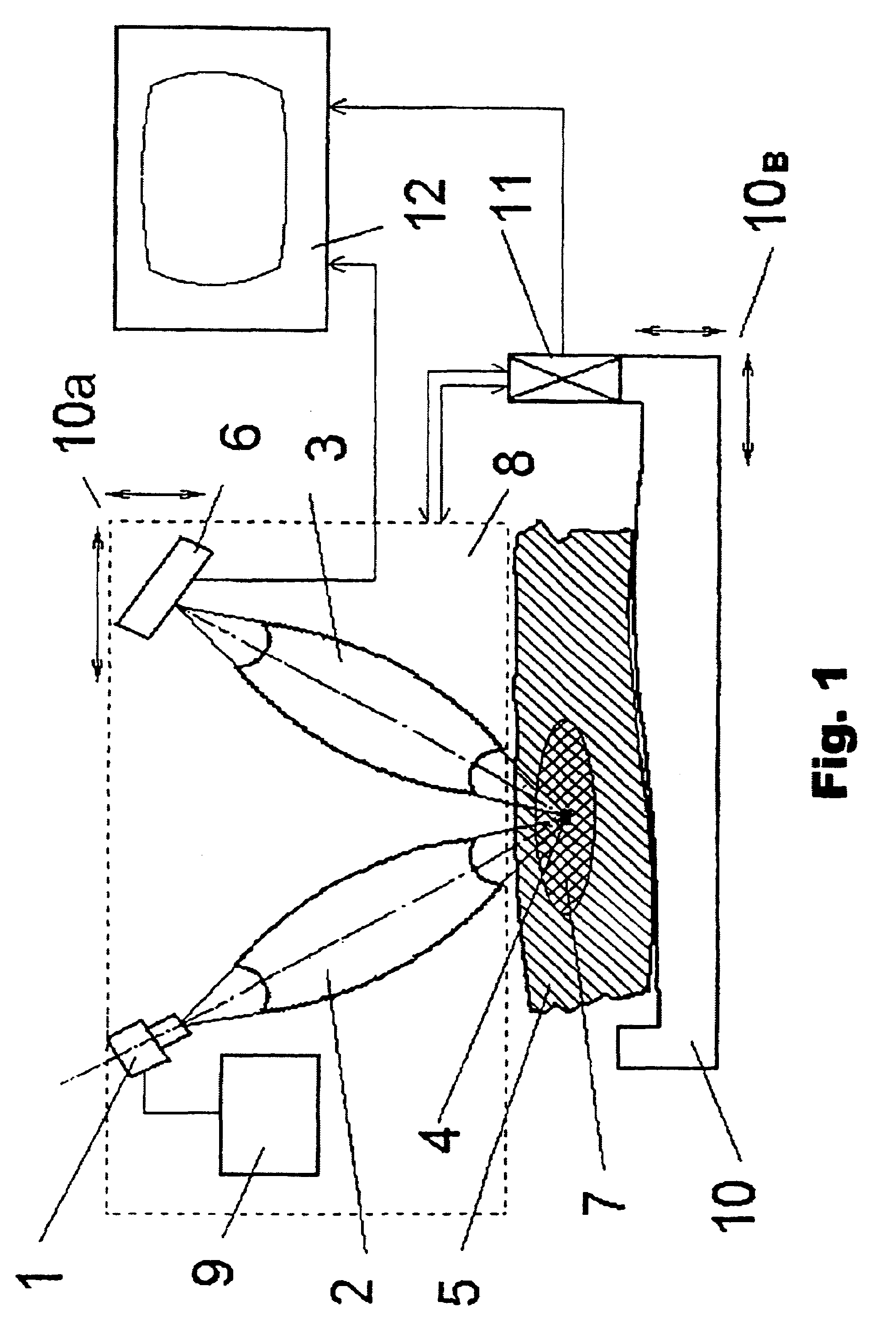
[0065]Divergent X-ray radiation from quasi-point source 1, generating radiation with energy corresponding to...


PUM


Abstract

The methods comprise detection of gadolinium in human tissues and organs by refining the position of malignant neoplasm and radiation treatment of the neoplasm in order to damage its cells. Refined position of the malignant neoplasm is determined by introducing gadolinium into the patient body and scanning part of the patient body in which the neoplasm is located. The scanning is performed by displacement of zone of radiation concentration created by intersection of several X-ray beams having radiation energy corresponding to K-edge of gadolinium atoms absorption. The refined information is acquired using detectors sensitive to Kα-radiation of gadolinium atoms, to which secondary radiation is transported arising in zone of concentration. Scanning is performed of the region of the malignant neoplasm location utilizing the same means as in the first stage. Sources of X-ray radiation are switched with control means into elevated intensity mode, sufficient for radiation damage of the malignant neoplasm tissues.

Description

FIELD OF THE INVENTION[0001]The invention relates to the means for position determination of malignant neoplasms in the patient body and their exposure with the purpose of malignant neoplasm cells injury to X-ray radiation.BACKGROUND OF THE INVENTION[0002]Methods are known which provide for carrying out the preliminary preparation after diagnosis being established and decision made on radiation therapy application to the malignant neoplasm with the purpose of damaging its cells. In the course of preliminary preparation, linear dimensions, area and volume of pathological masses, organs and anatomic structures is determined and their positional relationship is described in quantitative terms for particular patient (see, for example: Radiation therapy of malignant tumors. Physician's handbook. Prof. E. S. Kiseleva, Ed., Moscow, “Meditsina” publishing house, 1996 [1], p.46-47). Principal task of the preliminary preparation lies in merging different data obtained in the course of the dis...

Claims


Application Information

Patent Timeline
Patent Type & Authority Patents(United States)
IPC IPC(8): A61B6/00A61B5/05A61N5/10
CPCA61B6/4007A61B6/504A61N5/1084A61B6/481A61N5/1049A61N2005/109
Inventor KUMAKHOV, MURADIN ABUBEKIROVICH
Owner KUMAKHOV MURADIN ABUBEKIROVICH
Learn More