
Compositions for use in identification of viral hemorrhagic fever viruses

a technology for viral hemorrhagic fever and compositions, applied in the field of genetic identification and quantification of viruses, can solve the problems of rapid development of overwhelming viremia, fever, malaise, edema and hypotension,

Inactive Publication Date: 2007-12-25
IBIS BIOSCI
View PDF2 Cites 112 Cited by
  • Summary
  • Abstract
  • Description
  • Claims
  • Application Information

AI Technical Summary

Benefits of technology

Enables rapid, specific identification of unknown viruses without sequencing, providing a broad-range detection method that can characterize bioagents at the sub-species level, improving response times in bioterrorism and public health emergencies.

Problems solved by technology

These highly infectious viruses lead to a potentially lethal disease syndrome characterized by fever, malaise, vomiting, mucosal and gastrointestinal (GI) bleeding, edema and hypotension.
Inadequate or delayed immune response to these novel viral antigens may lead to rapid development of overwhelming viremia.
Ebola and Marburg viruses can cause severe hemorrhagic fever and have a high mortality rate.
LCMV has a world-wide geographic distribution and infection with LCMV leads to fever, malaise, weakness, myalgia and severe headache.
The remaining disease-causing arenaviruses are more limited in their distribution.
A problem in determining the cause of a natural infectious outbreak or a bioterrorist attack is the sheer variety of organisms that can cause human disease.
Although this approach is appropriate for the most obvious bioterrorist organisms, like smallpox and anthrax, experience has shown that it is very difficult to predict which of hundreds of possible pathogenic organisms might be employed in a terrorist attack.
The drawback of this approach for unknown bioagent detection and epidemiology is that analysis of the PCR products requires the cloning and sequencing of hundreds to thousands of colonies per sample, which is impractical to perform rapidly or on a large number of samples.
However, as with bacteria, there is no adequate analytical method other than sequencing to identify the viral bioagent present.
DNA chips with specific probes can only determine the presence or absence of specifically anticipated organisms.
Because there are hundreds of thousands of species of benign pathogens, some very similar in sequence to threat organisms, even arrays with 10,000 probes lack the breadth needed to identify a particular organism.

Method used


Image

  • Compositions for use in identification of viral hemorrhagic fever viruses
  • Compositions for use in identification of viral hemorrhagic fever viruses
  • Compositions for use in identification of viral hemorrhagic fever viruses

Examples


example 1

Selection of Primers that Define Bioagent Identifying Amplicons for VHF Viruses

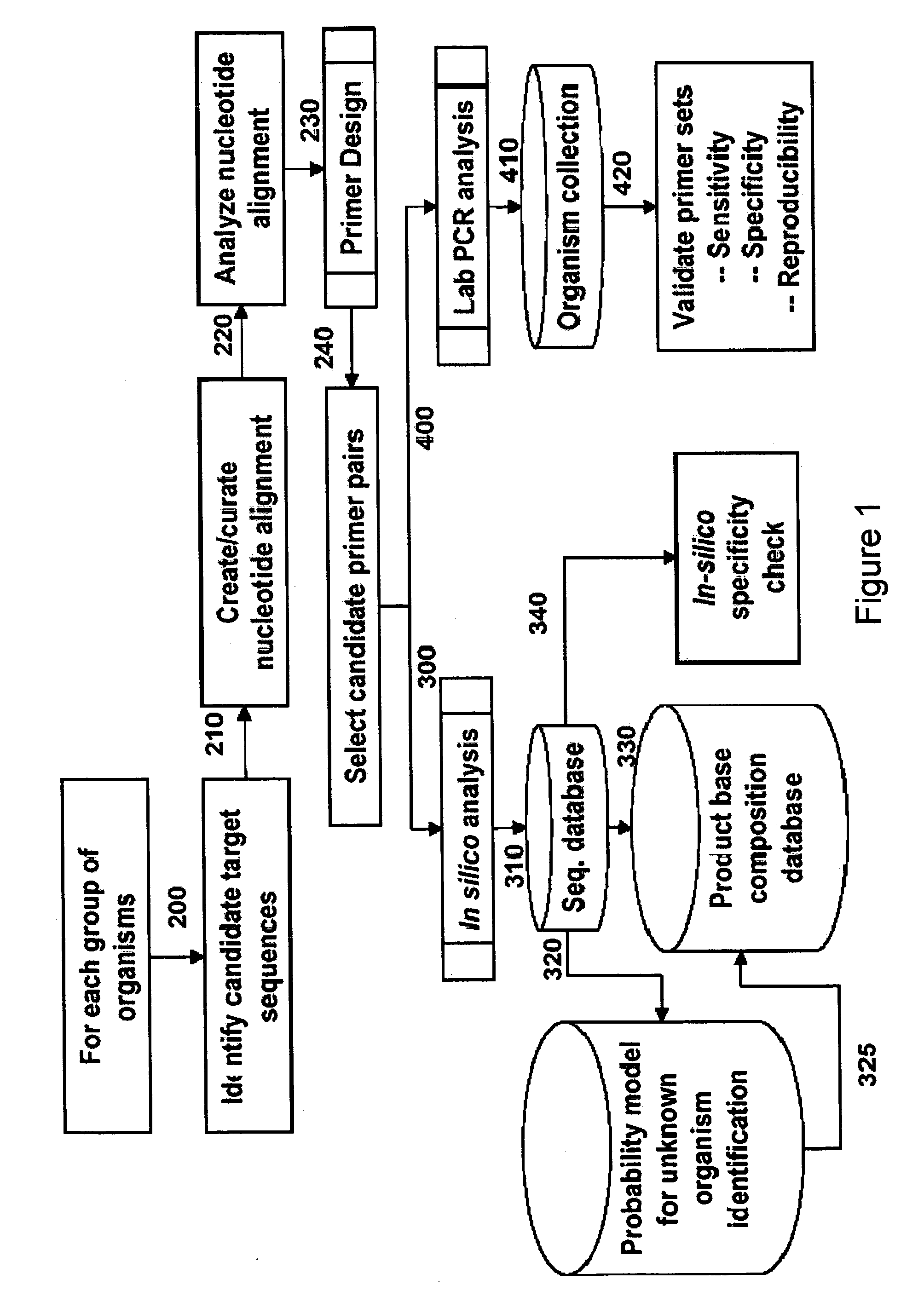
[0192]For design of primers that define viral hemorrhagic fever virus bioagent identifying amplicons, relevant sequences from, for example, GenBank were obtained, aligned and scanned for regions where pairs of PCR primers would amplify products of about 45 to about 200 nucleotides in length and distinguish species and / or sub-species from each other by their molecular masses or base compositions. A typical process shown in FIG. 1 is employed.

[0193]A database of expected base compositions for each primer region is generated using an in silico PCR search algorithm, such as (ePCR). An existing RNA structure search algorithm (Macke et al., Nucl. Acids Res., 2001, 29, 4724-4735, which is incorporated herein by reference in its entirety) has been modified to include PCR parameters such as hybridization conditions, mismatches, and thermodynamic calculations (SantaLucia, Proc. Natl. Acad. Sci. U.S.A., 1998, 95, 14...

example 2

One-Step RT-PCR of RNA Virus Samples

[0203]RNA was isolated from virus-containing samples according to methods well known in the art. To generate bioagent identifying amplicons for RNA viruses, a one-step RT-PCR protocol was developed. All RT-PCR reactions were assembled in 50 μl reactions in the 96 well microtiter plate format using a Packard MPII liquid handling robotic platform and MJ Dyad® thermocyclers (MJ research, Waltham, Mass.). The RT-PCR reaction consisted of 4 units of Amplitaq Gold®, 1.5× buffer II (Applied Biosystems, Foster City, Calif.), 1.5 mM MgCl2, 0.4 M betaine, 10 mM DTT, 20 mM sorbitol, 50 ng random primers (Invitrogen, Carlsbad, Calif.), 1.2 units Superasin (Ambion, Austin, Tex.), 100 ng polyA DNA, 2 units Superscript III (Invitrogen, Carlsbad, Calif.), 400 ng T4 Gene 32 Protein (Roche Applied Science, Indianapolis, Ind.), 800 μM dNTP mix, and 250 nM of each primer.

[0204]The following RT-PCR conditions were used to amplify the sequences used for mass spectromet...

example 3

Solution Capture Purification of PCR Products for Mass Spectrometry with Ion Exchange Resin-Magnetic Beads

[0205]For solution capture of nucleic acids with ion exchange resin linked to magnetic beads, 25 μl of a 2.5 mg / mL suspension of BioClon amine terminated supraparamagnetic beads were added to 25 to 50 μl of a PCR (or RT-PCR) reaction containing approximately 10 pM of a typical PCR amplification product. The above suspension was mixed for approximately 5 minutes by vortexing or pipetting, after which the liquid was removed after using a magnetic separator. The beads containing bound PCR amplification product were then washed 3× with 50 mM ammonium bicarbonate / 50% MeOH or 100 mM ammonium bicarbonate / 50% MeOH, followed by three more washes with 50% MeOH. The bound PCR amplicon was eluted with 25 mM piperidine, 25 mM imidazole, 35% MeOH, plus peptide calibration standards.


PUM

PropertyMeasurementUnit
molecular weightaaaaaaaaaa
molecular weightsaaaaaaaaaa
temperatureaaaaaaaaaa

Abstract

The present invention provides oligonucleotide primers, compositions, and kits containing the same for rapid identification of viruses that cause viral hemorrhagic fevers by amplification of a segment of viral nucleic acid followed by molecular mass analysis.

Description

CROSS REFERENCE TO RELATED APPLICATIONS [0001]This application claims the benefit of priority to U.S. Provisional Application Ser. No. 60 / 555,520 filed Mar. 22, 2004, which is incorporated herein by reference in its entirety.STATEMENT OF GOVERNMENT SUPPORT[0002]This invention was made with United States Government support under DARPA / SPO contract BAA00-09. The United States Government may have certain rights in the invention.FIELD OF THE INVENTION[0003]The present invention relates generally to the field of genetic identification and quantification of viruses in the Filoviridae, Flaviviridae, Bunyaviridae and Arenaviridae families and provides methods, compositions and kits useful for this purpose, as well as others, when combined with molecular mass analysis.BACKGROUND OF THE INVENTIONA. Viral Hemorrhagic Fever[0004]Viral hemorrhagic fevers (VHFs) are a group of febrile illnesses caused by RNA viruses from several viral families. These highly infectious viruses lead to a potentiall...

Claims


Application Information

Patent Timeline
Patent Type & Authority Patents(United States)
IPC IPC(8): C12Q1/68C12N15/11C12N15/33C12N15/40C12P19/34C12Q1/70
CPCC12Q1/70
Inventor SAMPATH, RANGARAJANHALL, THOMAS A.ESHOO, MARK W.
Owner IBIS BIOSCI
Learn More