
Method and system for simultaneously imaging multiple views of a plant embryo

a plant embryo and simultaneous imaging technology, applied in the field of simultaneous imaging of plant embryos, can solve the problems of time-consuming and expensive manual labor, high cost, and large amount of handling, and achieve the effect of shortening time and operation

Active Publication Date: 2007-10-30
WEYERHAEUSER NR CO
View PDF16 Cites 15 Cited by
  • Summary
  • Abstract
  • Description
  • Claims
  • Application Information

AI Technical Summary

Benefits of technology

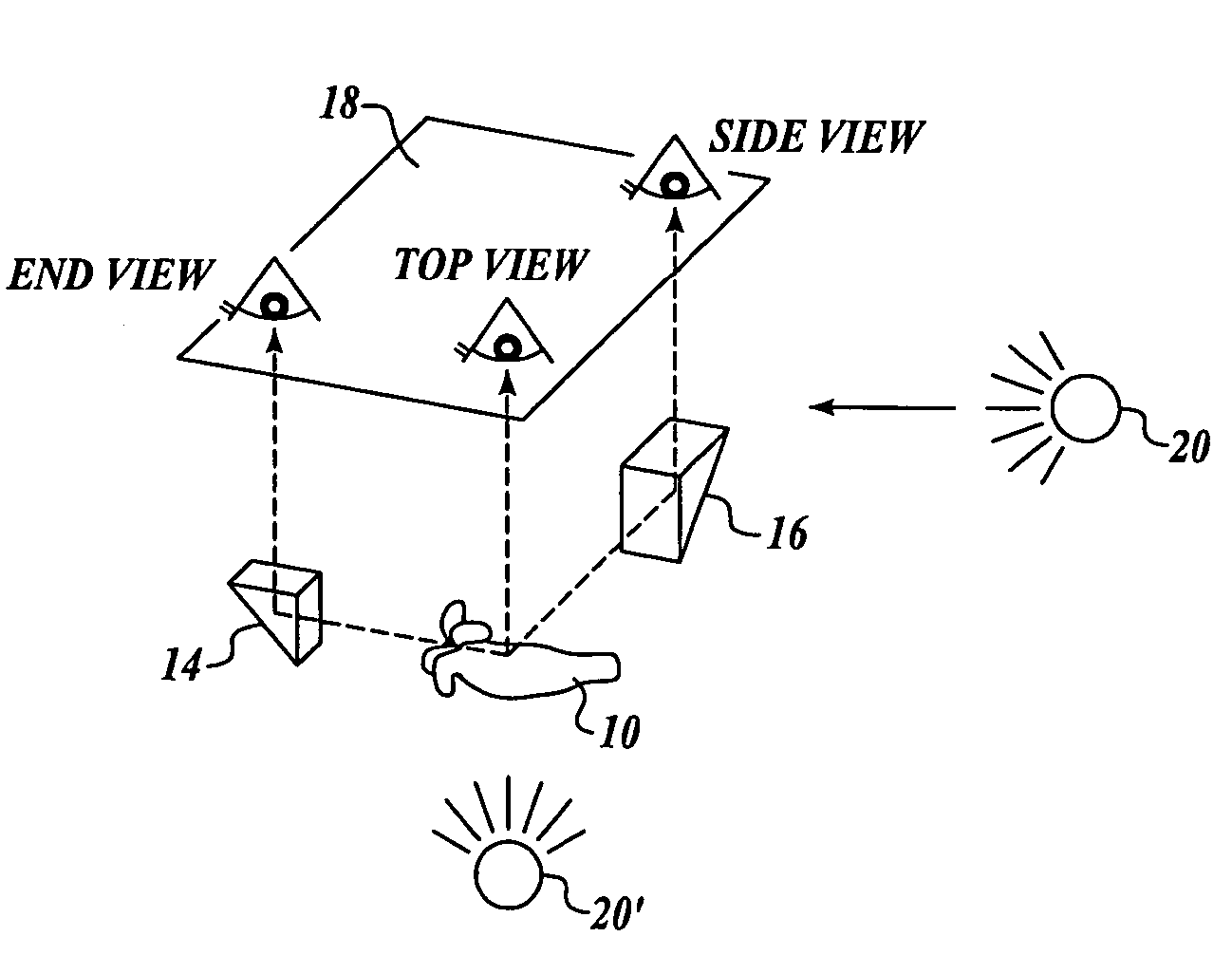
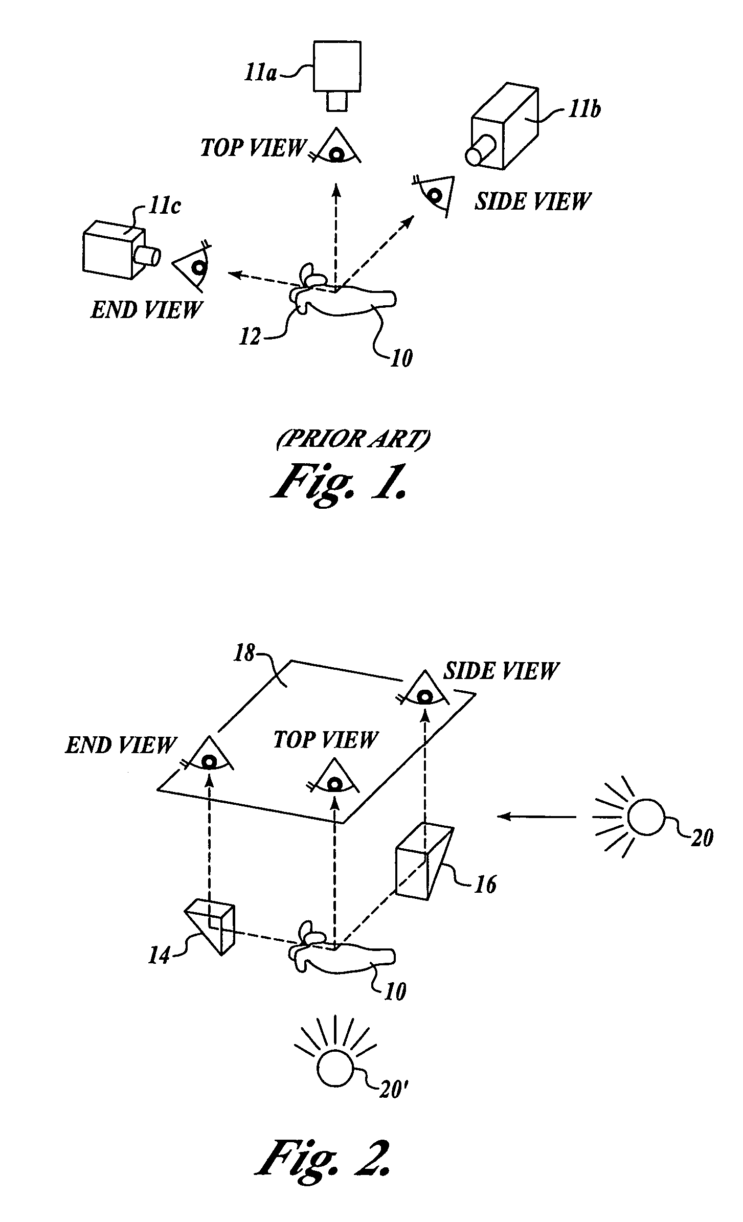
[0008]The invention provides a method of simultaneously imaging multiple views of a plant embryo. First, the method provides a camera for receiving a first view of a plant embryo (e.g., the top view). Second, the method provides a first reflecting surface for receiving and reflecting a second view of the plant embryo (e.g., the side view) toward the camera. Thus, using the camera, the method permits simultaneously imaging both the first and second views of the plant embryo. In one embodiment, the method further provides a second reflecting surface for receiving and reflecting a third view (e.g., the end view) of the plant embryo toward the camera, so that the camera can simultaneously image the first, second, and third views of the plant embryo.
[0012]According to yet another aspect of the invention, a light source is provided adjacent to the plant embryo to illuminate the plant embryo during image acquisition. Further, a cube-like enclosure, the interior surface of which is advantageously a white diffuse reflecting surface, may be provided to provide for diffuse lighting (substantially uniform illumination from all directions). The diffuse lighting arrangement eliminates shadows and prevents specular reflections from any wet or shiny areas on the surface of the embryo or from the interior surface of the enclosure. The enclosure also prevents undesirable light from reaching the image sensor of the camera. The enclosure including its various components is referred to as an “imaging cube.”
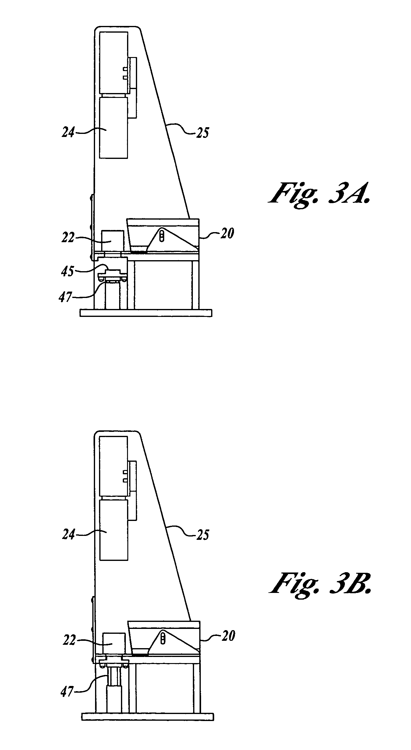
[0014]According to one aspect of the invention, the system of the present invention further includes a cube-like enclosure that is configured to substantially enclose the first and second reflecting surfaces and the embryo to be imaged, so as to provide diffuse lighting and to prevent undesirable light from reaching the image sensor of the camera. In one embodiment, the cube includes generally three openings: first opening to transmit the multiple views of the embryo to the camera; a second opening for receiving light from a light source to illuminate the plant embryo inside the cube during image acquisition; and a third opening for receiving the plant embryo to be viewed, perhaps as placed on an elevatable platform. In operation, the platform is lowered and a plant embryo to be viewed is placed thereon. Thereafter, the platform carrying the embryo is elevated to be received within the third opening of the cube, so as to substantially enclose the embryo to be imaged within the cube.
[0016]As will be apparent to those skilled in the art, the present invention offers several significant advantages over the previous method of taking images of an embryo using three orthogonally arranged cameras. First, there is no need to orient the embryo when taking one image relative to the orientation of the other two images, since three views of the embryo are taken simultaneously (with their relative orientation being fixed due to the fixed relationship of the camera and the reflecting surfaces). Second, the time required to send the three views of an embryo to a computer for analysis is cut by two thirds because, according to the invention, a single image combining all three views of an embryo can be transmitted to the computer. Thus, the present invention substantially shortens the time and operation required to obtain multiple views of an embryo, which can then be used to classify the embryos based on their desirability (or germination vigor).

Problems solved by technology

While conifer organogenesis was a breakthrough, costs were high due to the large amount of handling needed.
One of the more labor intensive and subjective steps in the embryogenesis procedure is the selection of individual embryos suitable for germination (e.g., incorporation into manufactured seeds).
This is a skilled yet tedious manual labor that is time consuming and expensive.
Further, it poses a major production bottleneck when the ultimate desired output will be in the millions of plants.
While the instrumental imaging analysis could replace the costly manual labor required to classify embryos based on their desirability, mass classification of embryos, in particular for the purpose of mass production of manufactured seeds, would require further shortening of time and lessening of operation required to classify embryos.

Method used


Image

  • Method and system for simultaneously imaging multiple views of a plant embryo
  • Method and system for simultaneously imaging multiple views of a plant embryo
  • Method and system for simultaneously imaging multiple views of a plant embryo

Examples


Embodiment Construction

[0025]FIG. 2 schematically illustrates the method of the present invention for simultaneously imaging multiple views of a plant embryo. The method involves directly imaging a first view of a plant embryo 10 (the top view in FIG. 2) on an image plane 18 of a camera; and using a first reflecting surface 14 to receive and reflect a second view of the embryo (the cotyledon end view in FIG. 2) toward the image plane 18 of the same camera. Thus, an image combining both the first and second views (i.e., the top and end views in the illustrated embodiment) can be taken. In one embodiment, the method further involves using a second reflecting surface 16 to receive and reflect a third view of the embryo (the side view in FIG. 2) toward the image plane 18 of the same camera. According to this arrangement, a camera with the image plane 18 can simultaneously acquire the first view (e.g., the top view) directly from the embryo 10, the second view (e.g., the cotyledon end view) via the first refle...


PUM

PropertyMeasurementUnit
lengthaaaaaaaaaa
surface areaaaaaaaaaaa
surface textureaaaaaaaaaa

Abstract

The invention provides a method and system for simultaneously imaging multiple views of a plant embryo. First, the method provides a camera for receiving a first view of a plant embryo (e.g., the top view). Second, the method provides a first reflecting surface for receiving and reflecting a second view of the plant embryo (e.g., the side view) toward the camera. Thus, the method permits simultaneously imaging both the first and second views of the plant embryo. In one embodiment, the method further provides a second reflecting surface for receiving and reflecting a third view (e.g., the end view) of the plant embryo toward the camera, so that the camera can simultaneously image the first, second, and third views of the plant embryo. The invention reduces the time required to obtain multiple views of an embryo, which can then be analyzed to classify the embryos according to their germination vigor.

Description

FIELD OF THE INVENTION[0001]The invention is directed to imaging plant embryos for determination of suitability for germination or other treatment, and more particularly, to simultaneously imaging multiple views of a plant embryo so as to permit efficient mass selection of plant embryos suitable for incorporation into manufactured seeds.BACKGROUND OF THE INVENTION[0002]Reproduction of selected plant varieties by tissue culture has been a commercial success for many years. The technique has enabled mass production of genetically identical selected ornamental plants, agricultural plants and forest species. The woody plants in this last group have perhaps posed the greatest challenges. Some success with conifers was achieved in the 1970s using organogenesis techniques wherein a bud, or other organ, was placed on a culture medium where it was ultimately replicated many times. The newly generated buds were placed on a different medium that induced root development. From there, the buds h...

Claims


Application Information

Patent Timeline
Patent Type & Authority Patents(United States)
IPC IPC(8): G06K9/00A01C1/00A01C1/02A01H4/00C12N5/04G01N21/01G01N21/84G02B21/18G02B27/02G02B27/14G03B15/00G03B35/00G03B35/10G03B35/18G06T15/00
CPCA01H4/006G01N21/84G02B27/022G01N2021/8466G01N2201/0634G01N2021/177
Inventor HIRAHARA, EDSPENCER, PAUL
Owner WEYERHAEUSER NR CO
Learn More