
Method for the preparation of purified HCV RNA by exosome separation

a technology of exosome separation and purification method, which is applied in the field of hepatitis c virus isolation method, can solve the problems of poor cell culture system, limited study of hcv life cycle, and considerable hinderance in hcv research, and achieves the effect of simple preparation

Inactive Publication Date: 2007-04-03
GRIFOLS WORLDWIDE OPERATIONS +1
View PDF7 Cites 193 Cited by
  • Summary
  • Abstract
  • Description
  • Claims
  • Application Information

AI Technical Summary

Benefits of technology

[0011]However, despite the identification of this receptor for HCV, there remains a great need for a method that would allow the simple preparation of the RNA genome of HCV. This would considerably facilitate the progress of research into the biology of this virus and would accelerate the design of therapeutic and diagnostic reagents that are effective in the treatment and prevention of disease associated with Hepatitis C infection.SUMMARY OF THE INVENTION
[0013]It has been discovered that in individuals infected with HCV, the virus binds to exosome particles in the plasma and is concentrated in these particles. The purification of these particles thus allows the simple preparation of significant quantities of HCV RNA without the need for complicated or expensive purification procedures.

Problems solved by technology

HCV research is hindered very considerably by the limited host range of the virus.
Study of HCV life cycle has also been limited by the lack of an efficient cell culture system.
Furthermore, attempts to purify HCV from biological materials such as plasma and liver have failed.

Method used


Image

  • Method for the preparation of purified HCV RNA by exosome separation
  • Method for the preparation of purified HCV RNA by exosome separation
  • Method for the preparation of purified HCV RNA by exosome separation

Examples


example 2

Preparation of HCV RNA

[0041]Given the results presented above, exosomes can now be isolated from plasma of HCV-infected patients enriched in HCV RNA. The experimental approach is as follows.

[0042]Plasma from HCV-infected human blood recovered after Ficoll separation is processed following the differential centrifugation protocol described above. The exosome-enriched supernatant collected from the centrifugation step at 10000×g is incubated overnight with anti-CD81-coated magnetic beads. Alternatively, after two clearing steps, HCV-infected plasma may be centrifuged at 20000×g before overnight incubation with anti-CD81-coated magnetic beads. Magnetic beads are then washed three times in 1% BSA in 50 mM Tris-HCl (pH 8.0), 1 mM EDTA, and 100 mM NaCl (TEN) buffer by magnetic separation and viral RNA are extracted with the Viral Extraction Kit (Qiagen). Quantitative RT-PCR for HCV RNA is performed as previously described (Pileri et al., 1998).

[0043]Alternative methods to capture exosomes...

example 3

Isolation of Exosomes from the Blood of HCV-Infected Patients

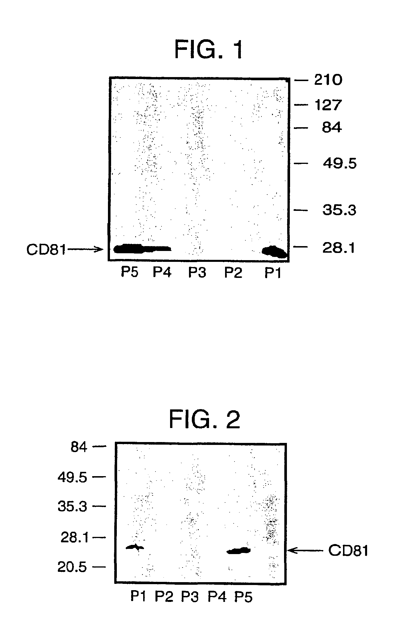
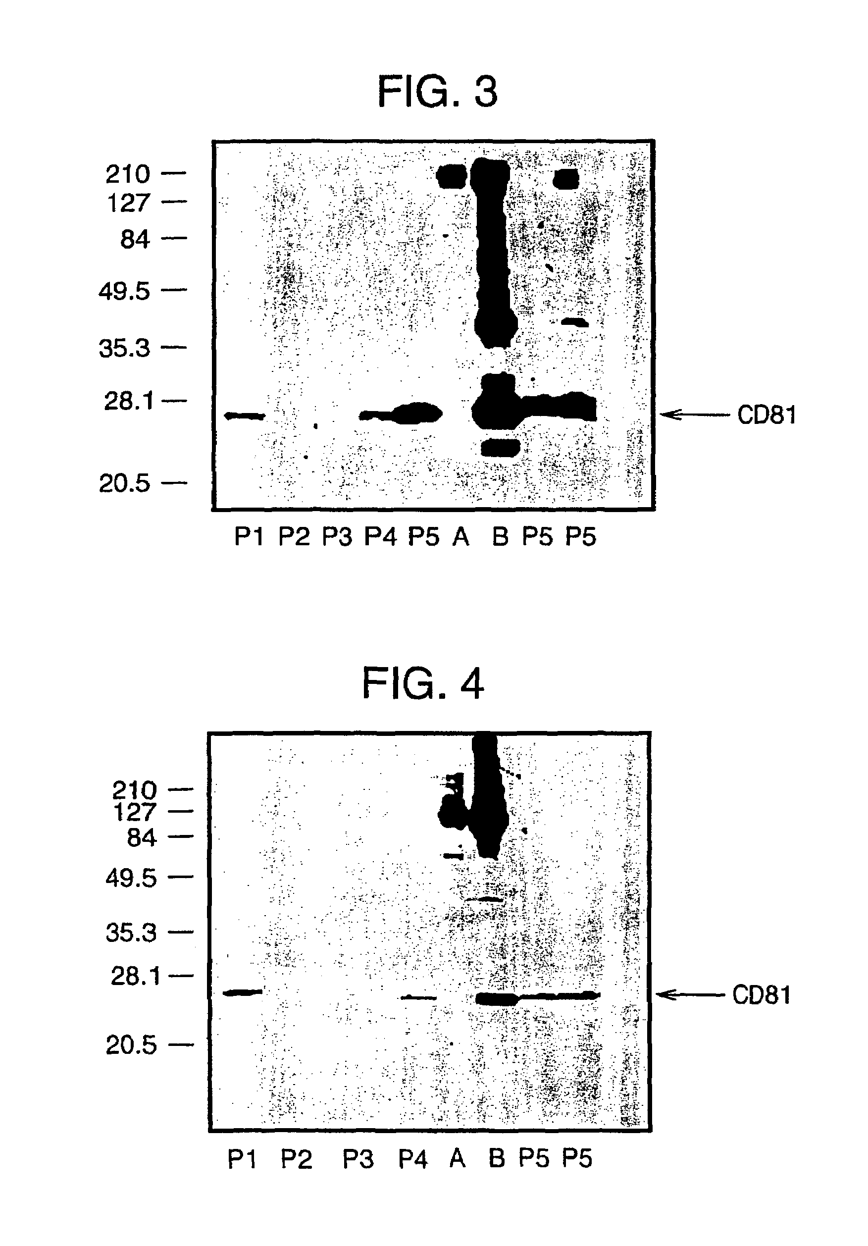
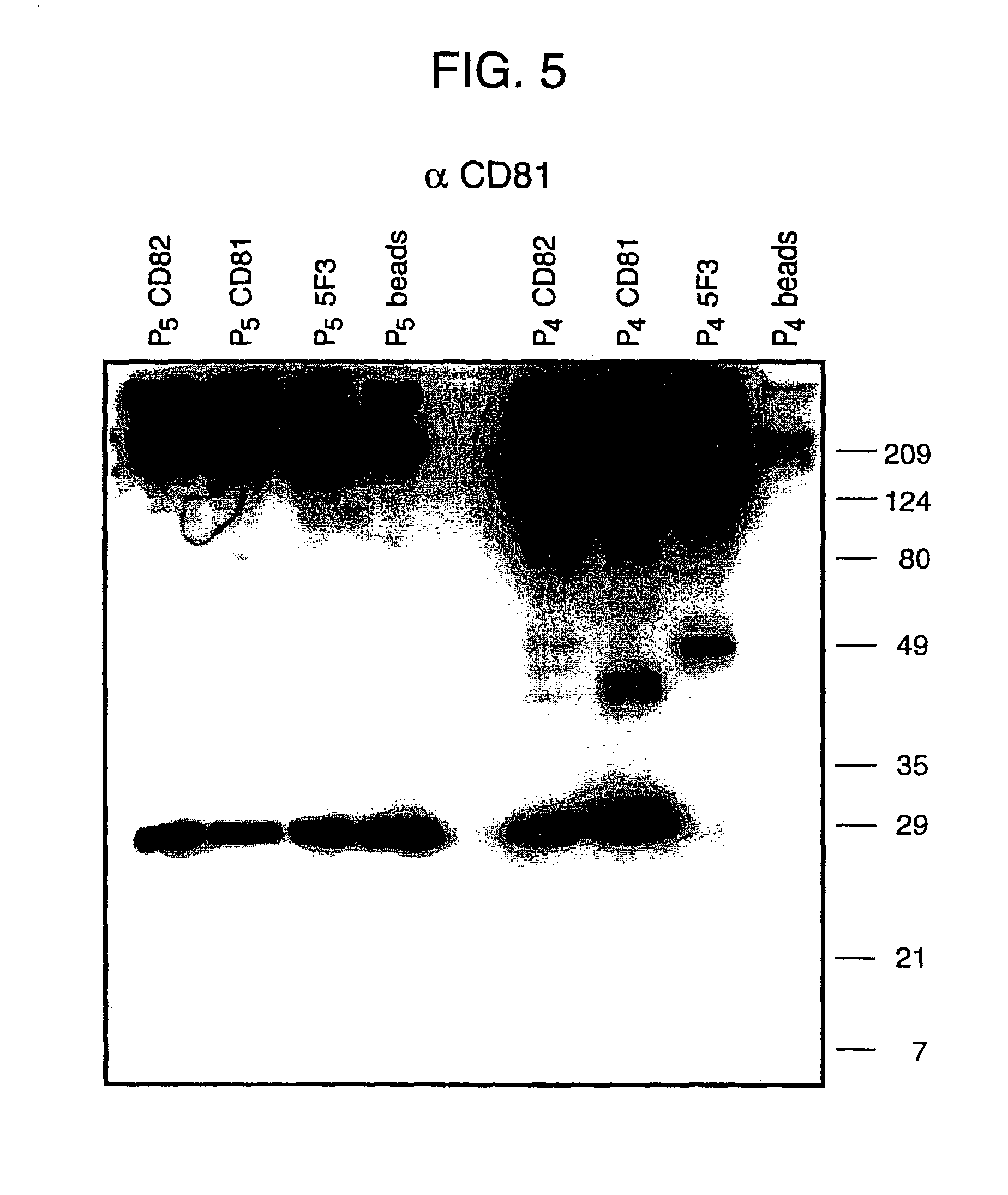
[0045]Here, we confirm the successful isolation of exosomes from the blood of HCV infected patients, either by steps of iterative centrifugation or by immunoselection with monoclonal antibodies against human CD81 or CD82 molecules, adopting the protocol used above for normal human plasma. Exosomes were then extracted in Laemmli buffer and CD81 (a marker enriched in exosomes) visualized by SDS-PAGE and Western blotting (see FIG. 5). This works confirms the presence of the CD81 protein in exosome preparations from the blood of an HCV patient.

[0046]Furthermore, in the exosomes prepared from infected patients, HCV RNA has been detected using quantitative RT PCR (see Table below).

[0047]Plasma from HCV-infected human blood recovered after Ficoll separation was processed following the differential centrifugation protocol described above. The exosome-enriched supernatant collected from the centrifugation step at 10000×g was incuba...


PUM

PropertyMeasurementUnit
pHaaaaaaaaaa
magneticaaaaaaaaaa
structureaaaaaaaaaa

Abstract

The invention relates to a method for the isolation of hepatitis C virus. The method comprises the separation of particles termed exosomes from the blood plasma of an individual infected with hepatitis C virus (HCV) and the extraction or RNA from these exosome particles.

Description

[0001]This application is a §371 filing from PCT / IB00 / 01801, filed Nov. 20, 2000, which claims priority from GB application 9927320.3, filed Nov. 18, 1999, from which applications priority is claimed pursuant to the provisions of 35 U.S.C. §§ 119 / 120 and which applications are incorporated by reference herein in their entireties.[0002]The present invention relates to a method for the isolation of hepatitis C virus. The method comprises the separation of particles termed exosomes from the blood plasma of an individual infected with hepatitis C virus (HCV) and extracting RNA from these exosome particles.[0003]All publications, manuals, patents, and patent applications cited herein are incorporated in full by reference.[0004]HCV (previously known as Non-A Non-B hepatitis—NANBV) is a positive sense RNA virus of about 10000 nucleotides with a single open reading frame encoding a polyprotein of about 3000 amino acids. Although the structure of the virus has been elucidated by recombinant ...

Claims


Application Information

Patent Timeline
Patent Type & Authority Patents(United States)
IPC IPC(8): C12P19/34C12Q1/70C12N15/09C07K14/18C12N7/02C12Q1/68C12R1/93
CPCC07K14/005C12N7/00A61K2039/525C12N2770/24222C12N2770/24251
Inventor ABRIGNANI, SERGIOPILERI, PIERO
Owner GRIFOLS WORLDWIDE OPERATIONS
Learn More