Methods and devices for promoting endothelial morphogenesis
a technology of endothelial morphogenesis and growth factors, which is applied in the direction of angiogenin, prosthesis, drug composition, etc., can solve the problems of affecting the function of implants, affecting the blood supply to a portion of heart muscles, and affecting the function of blood vessels, so as to reduce or prevent the formation of collagenous capsules, promote the growth of blood vessels, and reduce or prevent foreign body reactions
- Summary
- Abstract
- Description
- Claims
- Application Information
AI Technical Summary
Benefits of technology
Problems solved by technology
Method used
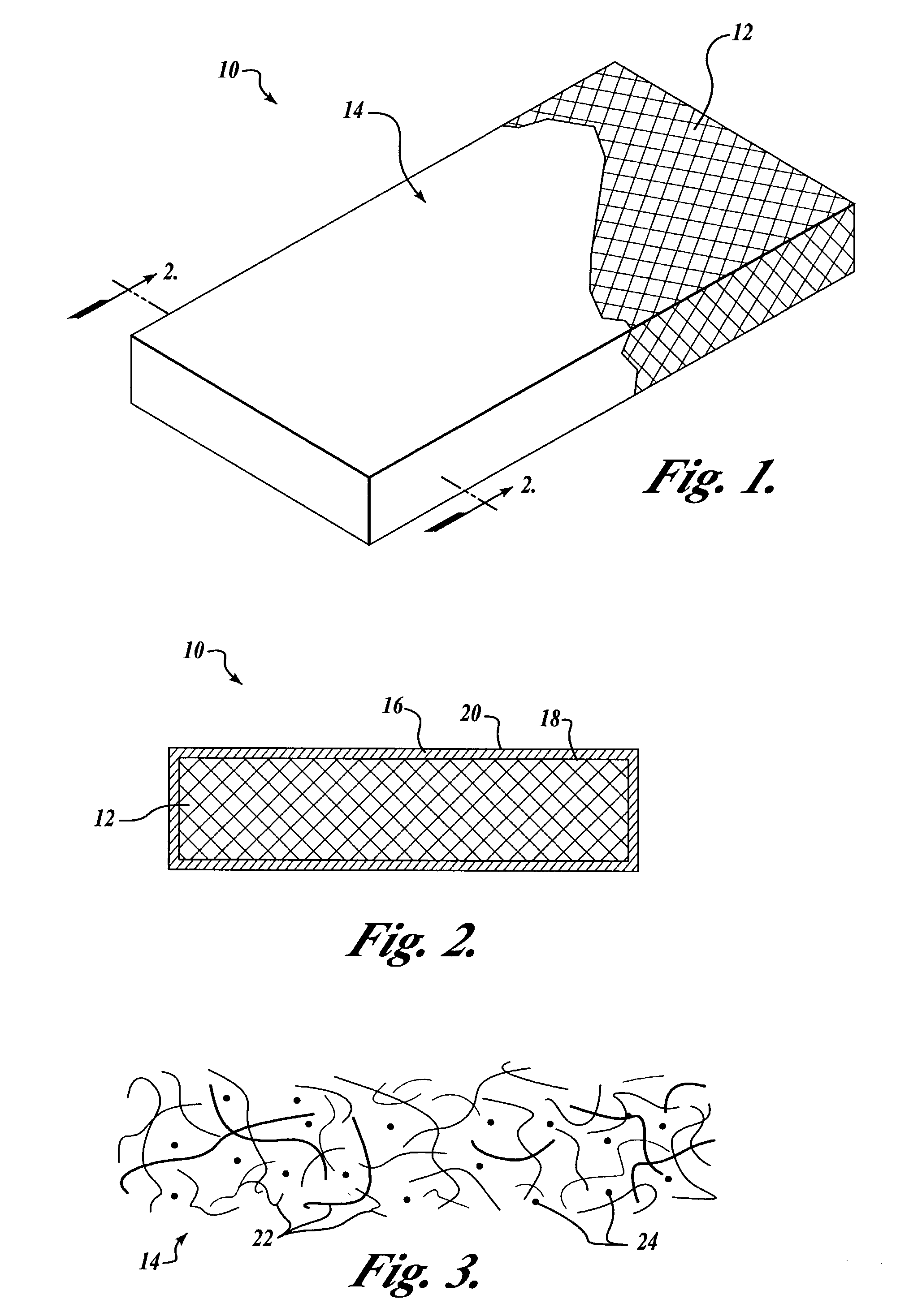
Image

Examples
example 1
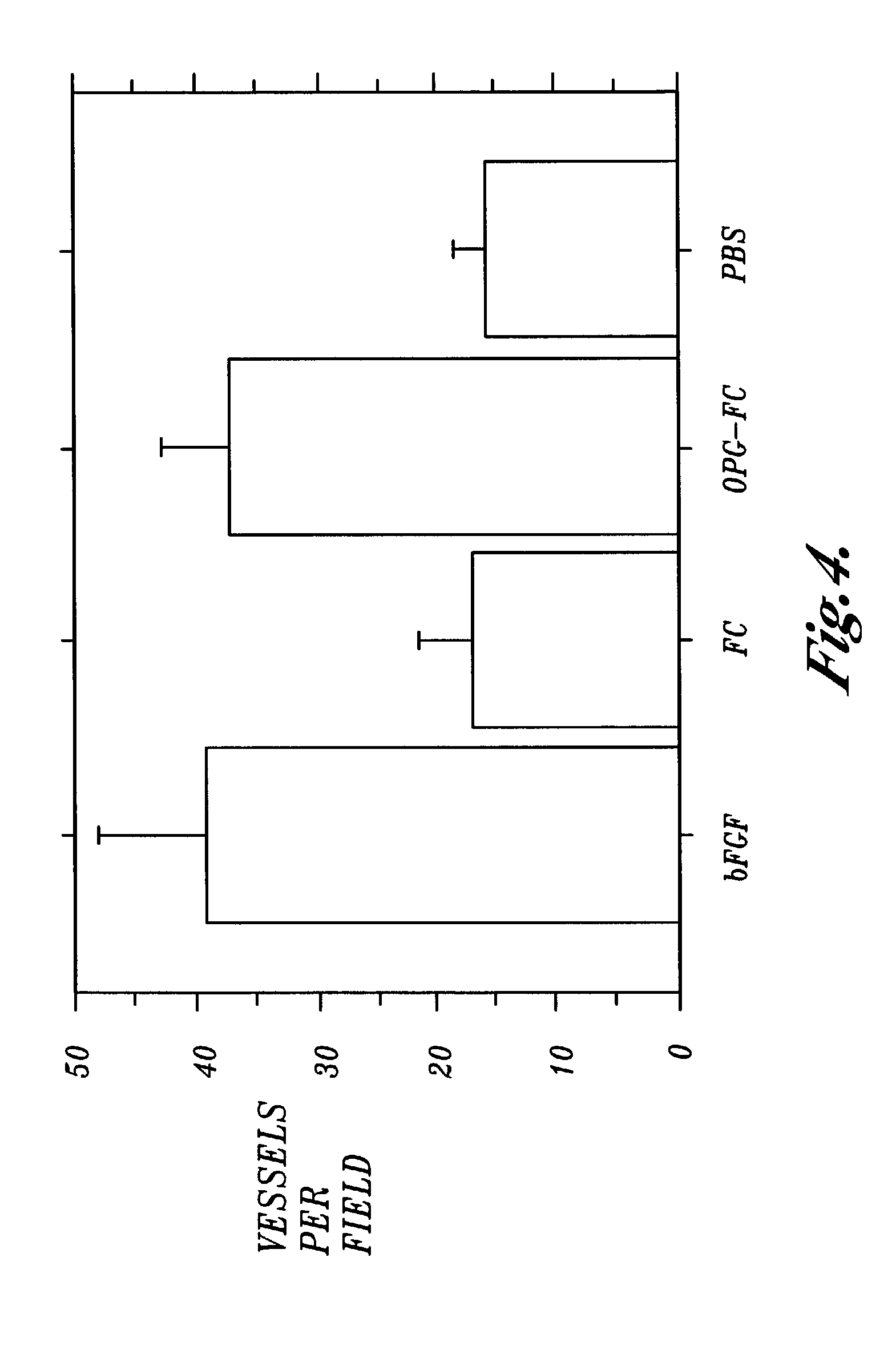
[0061]This Example shows that an osteoprotegerin-IgGFc protein fusion is as effective as fibroblast growth factor at inducing the formation of blood vessels in an in vivo impregnated sponge assay.
[0062]C57BL6 mice were implanted with polyvinyl alcohol sponges presoaked in one of the following solutions which each had a concentration of 100 ng / ml: a solution of bovine fibroblast growth factor (bFGF); a solution of the Fc portion of human IgG (FC); and a solution of a hybrid protein composed of the Fc portion of human IgG fused to osteoprotegerin (OPG-FC). The control was phosphate buffered saline (PBS).
[0063]After the sponges had been implanted subcutaneously in the backs of the mice for a period of fourteen days, the sponges were removed and the number of blood vessels growing into the sponges were counted under a microscope. The results are shown in FIG. 4, and show that osteoprotegerin-IgGFc protein fusion was as effective as fibroblast growth factor at inducing the formation of b...
example 2
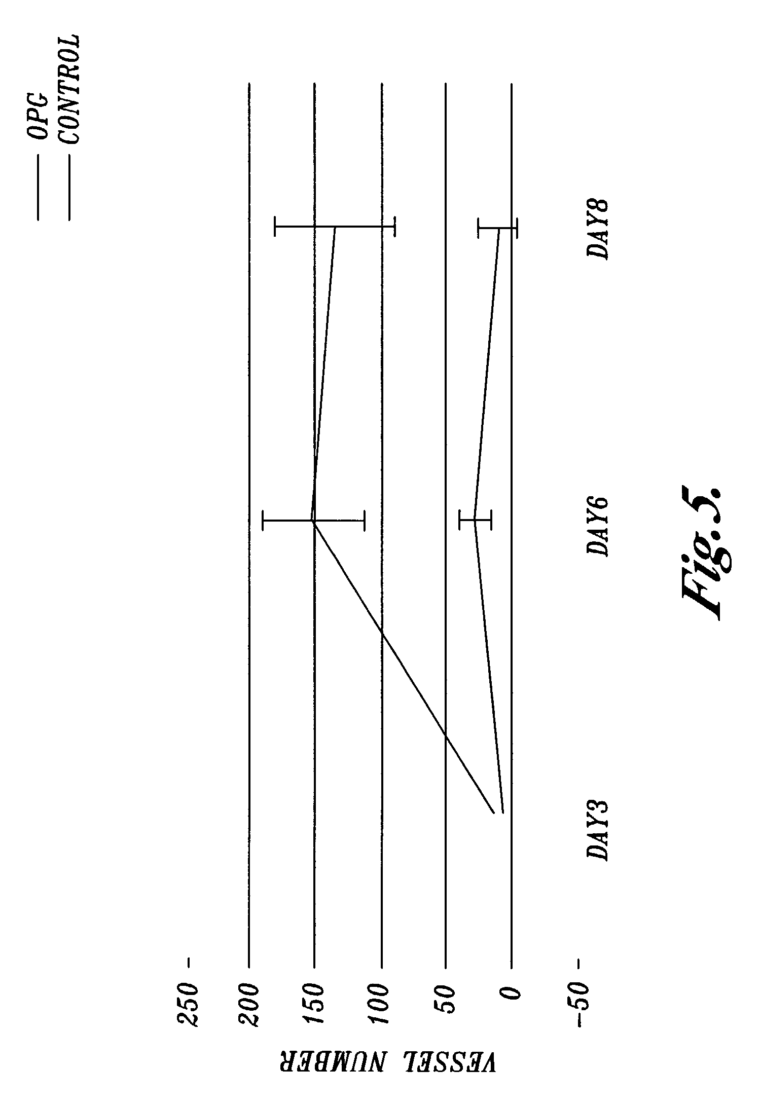
[0064]This Example shows that osteoprotegerin protein induces the formation of blood vessels in an in vitro rat aortic ring assay.
[0065]Thoracic aorta was excised from five to ten week old Fischer 344 male rats. The periaortic fibroadipose tissue was dissected and the cleaned aorta was cross-sectioned to yield rings of 1–2 mm in length. The rings were embedded in collagen gels. The collagen gels were prepared by mixing eight volumes of 1 mg / ml collagen with one volume of 10×Minimal Essential Medium (MEM, Invitrogen), pH 4.0, and one volume of 23.4 mg / ml NaHCO3. The embedded aortic rings were transferred to 16 mm wells that each contained 0.5 ml of serum-free endothelial basal medium (sold by Invitrogen as MCDB 131). The medium was changed three times per week starting at day three. The cultures were treated with a 140 nM solution of human recombinant osteoprotegerin. The control was aortic sections treated with Phosphate Buffered Saline only. The number of blood vessels growing from...
example 3
[0067]This example describes a representative hybridization protocol that can be used to identify nucleic acid molecules that encode an osteoprotegerin, or a portion of an osteoprotegerin that has the ability to promote endothelial morphogenesis, and that hybridize to the complement of the nucleic acid molecule consisting of the nucleic acid sequence set forth in SEQ ID NO:1, under defined hybridization conditions. In this Example, the complement of the nucleic acid molecule consisting of the nucleic acid sequence set forth in SEQ ID NO:1 is used as probe.
[0068]Hybridization solution should preferably be prepared and filtered through a 0.45-micron disposable cellulose acetate filter. The composition of the hybridization solution is 6×SSC, 5×Denhardt's reagent, 0.5% sodium dodecyl sulfate (SDS), 100 μg / ml denatured, fragmented salmon sperm DNA. The abbreviation “SSC” refers to a buffer used in nucleic acid hybridization solutions. One liter of the 20× (twenty times concentrate) stock...
PUM
| Property | Measurement | Unit |
|---|---|---|
| Fraction | aaaaa | aaaaa |
Abstract
Description
Claims
Application Information
 - R&D
- Intellectual Property
- Life Sciences
- Materials
- Tech Scout
- Unparalleled Data Quality
- Higher Quality Content
- 60% Fewer Hallucinations
Browse by: Latest US Patents, China's latest patents, Technical Efficacy Thesaurus, Application Domain, Technology Topic, Popular Technical Reports.
© 2025 PatSnap. All rights reserved.Legal|Privacy policy|Modern Slavery Act Transparency Statement|Sitemap|About US| Contact US: help@patsnap.com